Japanese
English
- 有料閲覧
PPV購入案内
1,320円 (1,200円+税10%) こちらは、1132ページから1134ページの文献です。購入には医書.jp本会員登録が必要です。
会員登録完了後に改めて上記「購入サイトへ」を選択してください。
または「購入サイトへ」選択後に「購入ログイン」、「新規会員登録」の順に進んでください。
会員登録について詳しくはをご確認ください。
- Abstract 文献概要
- 1ページ目 Look Inside
はじめに
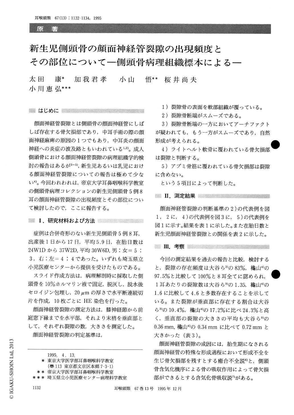
顔面神経管裂隙とは側頭骨の顔面神経管にしばしば存在する骨欠損部であり,中耳手術の際の顔面神経麻痺の原因の1つでもあり,中耳炎の顔面神経への炎症の波及路ともいわれている1,2)。成人側頭骨における顔面神経管裂隙の病理組織学的検討の報告はあるが3〜5),新生児あるいは乳児における顔面神経管裂隙についての報告は極めて少ない4)。今回われわれは,帝京大学耳鼻咽喉科学教室の側頭骨病理コレクションの新生児側頭骨5例8耳の顔面神経管裂隙の出現頻度とその部位について検討したので,ここに報告する。
The location and size of dehiscence of the facial canal were determined from histologic examination of 8 newborn temporal bones. Dihiscence was found in all temporal bones. A total of 37 dehiscences were found and the average number of dehiscence was 4.6 per of the temoral bones. The average width of dehiscence was 0.46 mm. 75.7% of the dehiscences were located in the horizontal segment and 24.3% in the vertical segment.
The average number of dehiscence per newborn temporal bone was higher and the percentage of dehiscence of the vertical segment of the newborn temporal bone was higher than that of adult. These findings support the theory that dehiscence appears due to incomplete fusion of the bone wall during the facial canal development process in the embryonal stage.
Copyright © 1995, Igaku-Shoin Ltd. All rights reserved.