Japanese
English
- 有料閲覧
PPV購入案内
1,320円 (1,200円+税10%) こちらは、267ページから270ページの文献です。購入には医書.jp本会員登録が必要です。
会員登録完了後に改めて上記「購入サイトへ」を選択してください。
または「購入サイトへ」選択後に「購入ログイン」、「新規会員登録」の順に進んでください。
会員登録について詳しくはをご確認ください。
- Abstract 文献概要
- 1ページ目 Look Inside
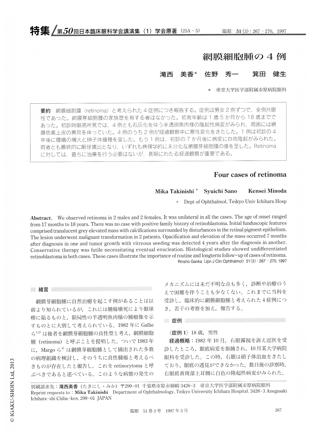
(25A-5) 網膜細胞腫(retinoma)と考えられた4症例につき報告する。症例は男女2例ずつで,全例片眼性であった。網膜芽細胞腫の家族歴を有する者はなかった。初発年齢は1歳5か月から18歳までであった。初診時眼底所見では,4例とも石灰化を伴う半透明魚肉様の隆起性病変がみられ,周囲には網膜色素上皮の異常を伴っていた。4例のうち2例が経過観察中に悪性変化をきたした。1例は初診の4年後に腫瘍の増大と硝子体播種を呈した。もう1例は,初診の7か月後に病変に白色隆起がみられた。両者とも最終的に眼球摘出となり,いずれも病理学的に未分化な網膜芽細胞腫の像を呈した。Retinomaに対しては,直ちに治療を行う必要はないが,長期にわたる経過観察が重要である。
We observed retinoma in 2 males and 2 females. It was unilateral in all the cases. The age of onset ranged from 17 months to 18 years. There was no case with positive family history of retinoblastoma. Initial funduscopic features comprised translucent grey elevated mass with calcifications surrounded by disturbances in the retinal pigment epithelium. The lesion underwent malignant transformation in 2 patients. Opacification and elevation of the mass occurred 7 months after diagnosis in one and tumor growth with vitreous seeding was detected 4 years after the diagnosis in another. Conservative therapy was futile necessitating eventual enucleation. Histological studies showed undifferentiated retinoblastoma in both cases. These cases illustrate the importance of routine and longterm follow-up of cases of retinoma.
Copyright © 1997, Igaku-Shoin Ltd. All rights reserved.