Japanese
English
- 有料閲覧
PPV購入案内
1,320円 (1,200円+税10%) こちらは、277ページから281ページの文献です。購入には医書.jp本会員登録が必要です。
会員登録完了後に改めて上記「購入サイトへ」を選択してください。
または「購入サイトへ」選択後に「購入ログイン」、「新規会員登録」の順に進んでください。
会員登録について詳しくはをご確認ください。
- Abstract 文献概要
- 1ページ目 Look Inside
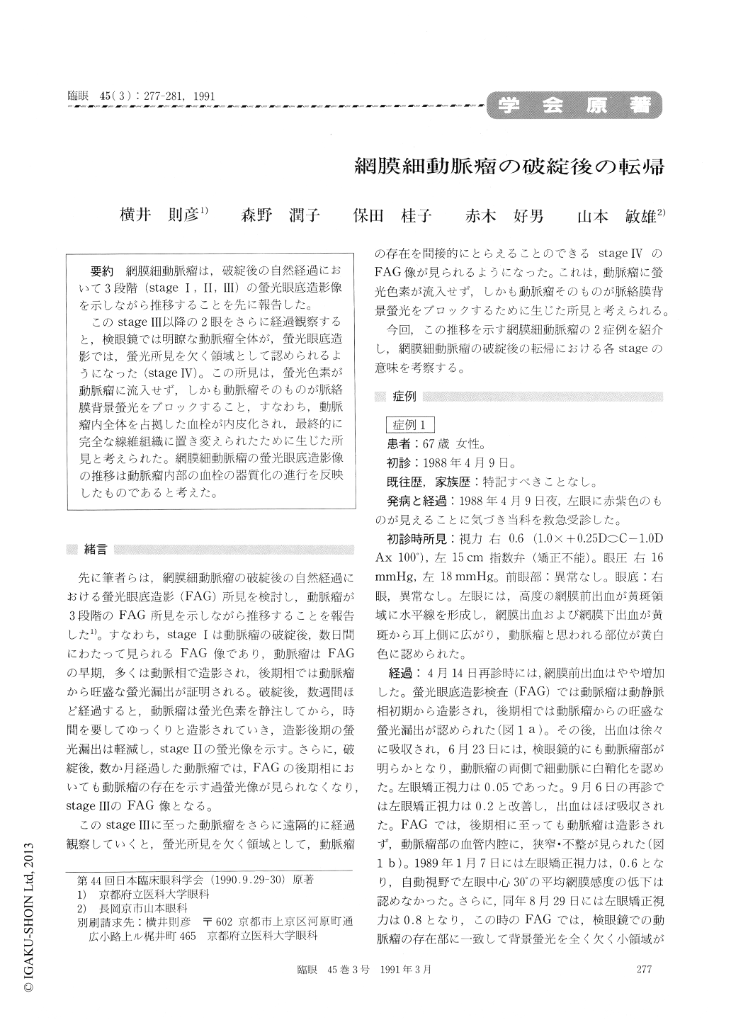
網膜細動脈瘤は,破綻後の自然経過において3段階(stageⅠ,Ⅱ,Ⅲ)の螢光眼底造影像を示しながら推移することを先に報告した。
このstageⅢ以降の2眼をさらに経過観察すると,検眼鏡では明瞭な動脈瘤全体が,螢光眼底造影では,螢光所見を欠く領域として認められるようになった(stageⅣ)。この所見は,螢光色素が動脈瘤に流入せず,しかも動脈瘤そのものが脈絡膜背景螢光をブロックすること,すなわち,動脈瘤内全体を占拠した血栓が内皮化され,最終的に完全な線維組織に置き変えられたために生じた所見と考えられた。網膜細動脈瘤の螢光眼底造影像の推移は動脈瘤内部の血栓の器質化の進行を反映したものであると考えた。
We observed, in our previous study, that ruptured retinal arterial macroaneurysm goes through 3 stages during the natural course of the disease. Immediately after rupture, the macroaneurysm shows prompt dye filling followed by massive extravasation (stageⅠ). A few weeks later, the macroaneurysm is slowly filled by the dye with less pronounced extravasation (stageⅡ). Months later,no trace of macroaneurysm in visible by fluorescein angiography (stageⅢ).
We observed the presence of stageⅣ in 2 eyes 14 and 16 months each after onset. Both eyes were characterized by the presence of small area of blocked fluorescence corresponding to the previous site of the macroaneurysm. The finding seemed to imply that the macroaneurysm had undergone com-plete fibrotic thrombosis.
Copyright © 1991, Igaku-Shoin Ltd. All rights reserved.