Japanese
English
- 有料閲覧
- Abstract 文献概要
- 1ページ目 Look Inside
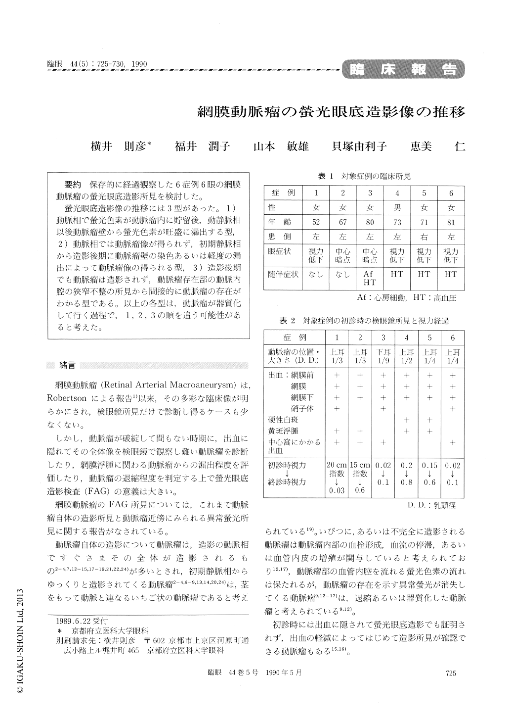
保存的に経過観察した6症例6眼の網膜動脈瘤の螢光眼底造影所見を検討した。
螢光眼底造影像の推移には3型があった。1)動脈相で螢光色素が動脈瘤内に貯留後,動静脈相以後動脈瘤壁から螢光色素が旺盛に漏出する型,2)動脈相では動脈瘤像が得られず,初期静脈相から造影後期に動脈瘤壁の染色あるいは軽度の漏出によって動脈瘤像の得られる型,3)造影後期でも動脈瘤は造影されず,動脈瘤存在部の動脈内腔の狭窄不整の所見から間接的に動脈瘤の存在がわかる型である。以上の各型は,動脈瘤が器質化して行く過程で,1,2,3の順を追う可能性があると考えた。
We followed up 6 cases of retinal arteriolar macroaneurysm with repeated fluorescein angio-graphy. We could distinguish three different angio-graphic patterns of the disease.
In the early stage of the disease, dye pooling in the aneurysm is seen during the arterial phase, followed by extravasation in the later phase. A fewweeks after occurrence of aneurysmal rupture, the aneurysm is not visible during the arterial phase. The aneurysm becomes fluorescent due to dye staining of its wall. After the aneurysm has cicatricialized, the aneurysm appears as filling defect. Its presence can be inferred by narrowing and irregularity of the involved retinal artery.

Copyright © 1990, Igaku-Shoin Ltd. All rights reserved.


