Japanese
English
- 有料閲覧
PPV購入案内
1,320円 (1,200円+税10%) こちらは、975ページから978ページの文献です。購入には医書.jp本会員登録が必要です。
会員登録完了後に改めて上記「購入サイトへ」を選択してください。
または「購入サイトへ」選択後に「購入ログイン」、「新規会員登録」の順に進んでください。
会員登録について詳しくはをご確認ください。
- Abstract 文献概要
- 1ページ目 Look Inside
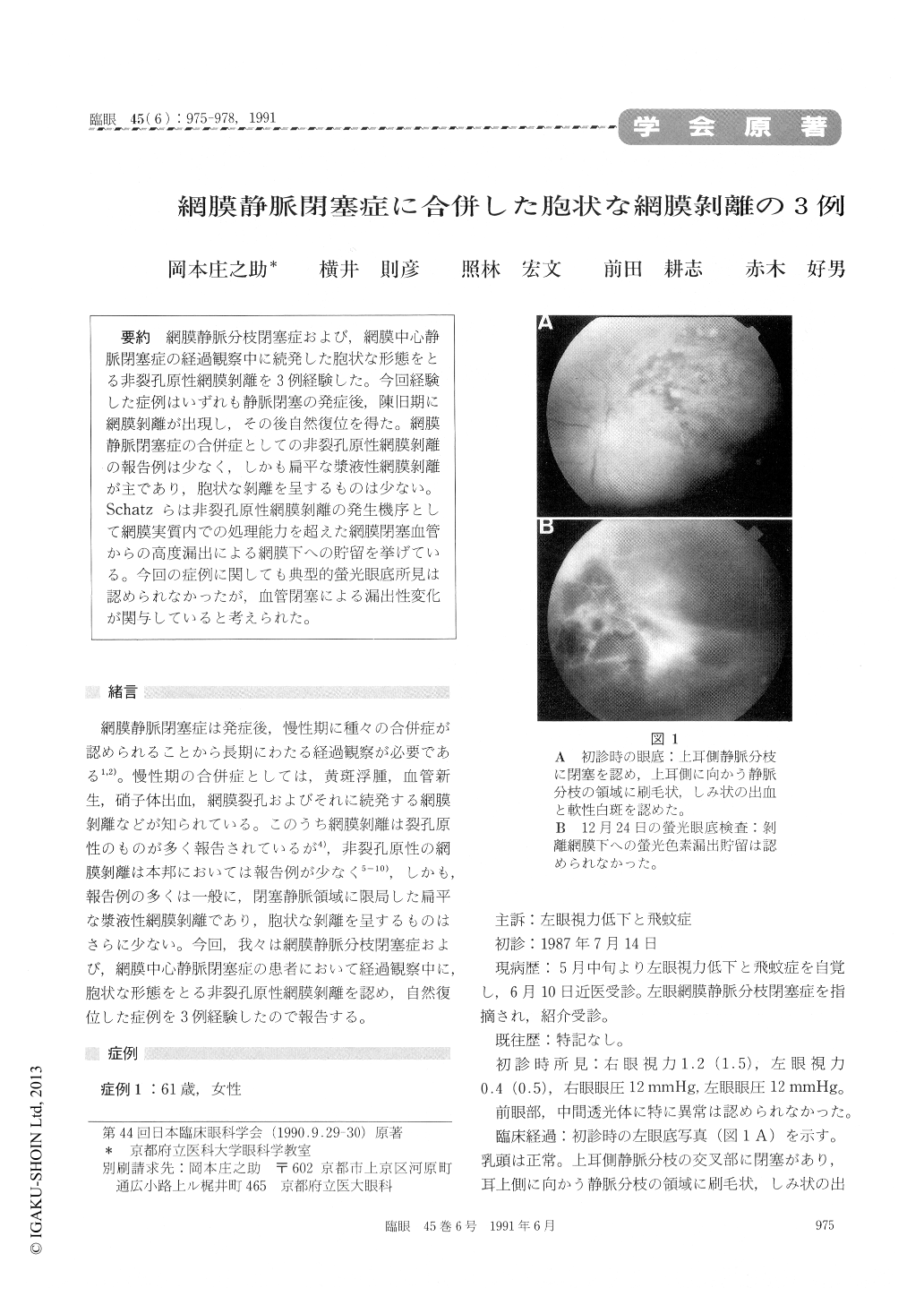
網膜静脈分枝閉塞症および,網膜中心静脈閉塞症の経過観察中に続発した胞状な形態をとる非裂孔原性網膜剥離を3例経験した。今回経験した症例はいずれも静脈閉塞の発症後,陳旧期に網膜剥離が出現し,その後自然復位を得た。網膜静脈閉塞症の合併症としての非裂孔原性網膜剥離の報告例は少なく,しかも扁平な漿液性網膜剥離が主であり,胞状な剥離を呈するものは少ない。Schatzらは非裂孔原性網膜剥離の発生機序として網膜実質内での処理能力を超えた網膜閉塞血管からの高度漏出による網膜下への貯留を挙げている。今回の症例に関しても典型的螢光眼底所見は認められなかったが,血管閉塞による漏出性変化が関与していると考えられた。
Three cases with retinal vein in its chronic stage occlusion developed nonrhegmatogenous retinal detachment. The detachment was bullous in all the eyes and was localized peripheral to the occluded vein. Fluorescein angiography disappeared was unremarkable except typical findings of retinal vein occlusion with capillary nonperfusion. Poolingof dye in the subretinal spke was consistently absent. The retinal detachment spontaneously disappeared in 2 to 5 weeks. The initially detached retinal area was then treated by photocoagulation. No recurrence of retinal detachment has been ob-served thereafter. It appeared that the bullous retinal detachment was secondary to transudation from affected retinal vessels.
Copyright © 1991, Igaku-Shoin Ltd. All rights reserved.