Japanese
English
- 有料閲覧
PPV購入案内
1,320円 (1,200円+税10%) こちらは、1129ページから1132ページの文献です。購入には医書.jp本会員登録が必要です。
会員登録完了後に改めて上記「購入サイトへ」を選択してください。
または「購入サイトへ」選択後に「購入ログイン」、「新規会員登録」の順に進んでください。
会員登録について詳しくはをご確認ください。
- Abstract 文献概要
- 1ページ目 Look Inside
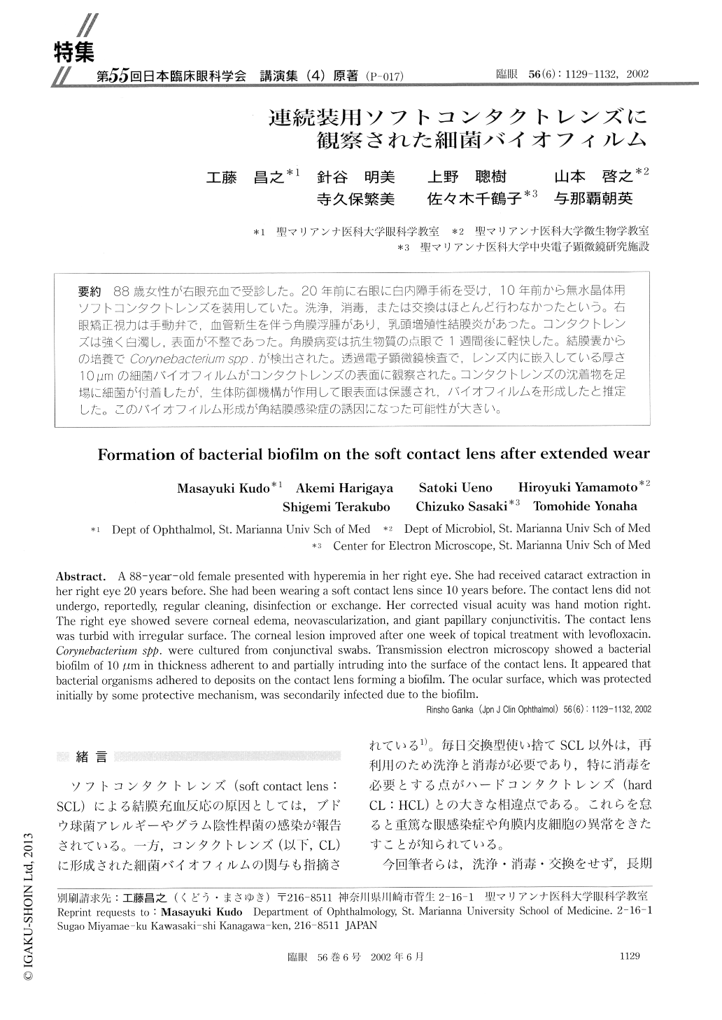
88歳女性が右眼充血で受診した。20年前に右眼に白内障手術を受け,10年前から無水晶体用ソフトコンタクトレンズを装用していた。洗浄,消毒,または交換はほとんど行わなかったという。右眼矯正視力は手動弁で、血管新生を伴う角膜浮腫があり,乳頭増殖性結膜炎があった。コンタクトレンズは強く白濁し,表面が不整であった。角膜病変は抗生物質の点眼で1週問後に軽快した。結膜嚢からの培養でCorynebacterium spp.が検出された。透過電子顕微鏡検査で,レンズ内に嵌入している厚さ10μmの細菌バイオフィルムがコンタクトレンズの表面に観察された。コンタクトレンズの沈着物を足場に細菌が付着したが、生体防御機構が作用して眼表面は保護され,バイオフィルムを形成したと推定した。このバイオフィルム形成が角結膜感染症の誘因になった可能性が大きい。
A 88-year-old female presented with hyperemia in her right eye. She had received cataract extraction in her right eye 20 years before. She had been wearing a soft contact lens since 10 years before. The contact lens did not undergo, reportedly, regular cleaning, disinfection or exchange. Her corrected visual acuity was hand motion right. The right eye showed severe corneal edema, neovascularization, and giant papillary conjunctivitis. The contact lens was turbid with irregular surface. The corneal lesion improved after one week of topical treatment with levofloxacin. Corynebacterium spp. were cultured from conjunctival swabs. Transmission electron microscopy showed a bacterial biofilm of 10 μm in thickness adherent to and partially intruding into the surface of the contact lens. It appeared that bacterial organisms adhered to deposits on the contact lens forming a biofilm. The ocular surface, which was protected initially by some protective mechanism, was secondarily infected due to the biofilm.
Copyright © 2002, Igaku-Shoin Ltd. All rights reserved.