Japanese
English
- 有料閲覧
PPV購入案内
1,320円 (1,200円+税10%) こちらは、489ページから492ページの文献です。購入には医書.jp本会員登録が必要です。
会員登録完了後に改めて上記「購入サイトへ」を選択してください。
または「購入サイトへ」選択後に「購入ログイン」、「新規会員登録」の順に進んでください。
会員登録について詳しくはをご確認ください。
- Abstract 文献概要
- 1ページ目 Look Inside
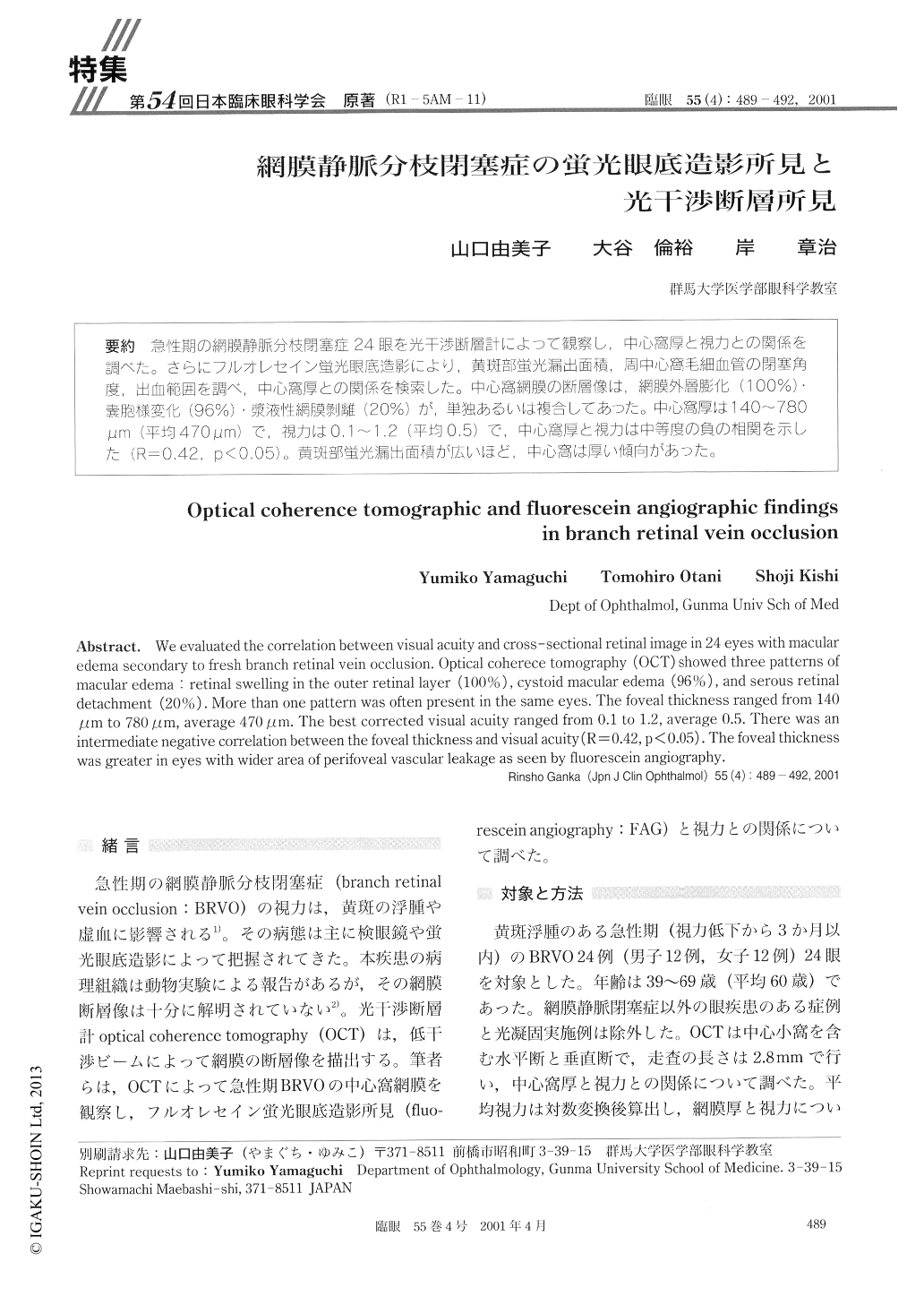
急性期の網膜静脈分枝閉塞症24眼を光干渉断層計によって観察し,中心窩厚と視力との関係を調べた。さらにフルオレセイン蛍光眼底造影により,黄斑部蛍光漏出面積,周中心窩毛細血管の閉塞角度,出血範囲を調べ,中心窩厚との関係を検索した。中心窩網膜の断層像は,網膜外層膨化(100%)・嚢胞様変化(96%)・漿液性網膜剥離(20%)が,単独あるいは複合してあった。中心窩厚は140〜780μm (平均470μm)で,視力はO.1〜1.2(平均0.5)で,中心窩厚と視力は中等度の負の相関を示した(R=0.42,p<0.05)。黄斑部蛍光漏出面積が広いほど、中心窩は厚い傾向があった。
We evaluated the correlation between visual acuity and cross-sectional retinal image in 24 eyes with macular edema secondary to fresh branch retinal vein occlusion. Optical coherece tomography (OCT) showed three patterns of macular edema : retinal swelling in the outer retinal layer (100%), cystoid macular edema (96%), and serous retinal detachment (20%). More than one pattern was often present in the same eyes. The foveal thickness ranged from 140 μm to 780μm, average 470μm. The best corrected visual acuity ranged from 0.1 to 1.2, average 0.5. There was an intermediate negative correlation between the foveal thickness and visual acuity (R=0.42, p<0.05). The foveal thickness was greater in eyes with wider area of perifoveal vascular leakage as seen by fluorescein angiography.
Copyright © 2001, Igaku-Shoin Ltd. All rights reserved.