Japanese
English
- 有料閲覧
PPV購入案内
1,320円 (1,200円+税10%) こちらは、1483ページから1488ページの文献です。購入には医書.jp本会員登録が必要です。
会員登録完了後に改めて上記「購入サイトへ」を選択してください。
または「購入サイトへ」選択後に「購入ログイン」、「新規会員登録」の順に進んでください。
会員登録について詳しくはをご確認ください。
- Abstract 文献概要
- 1ページ目 Look Inside
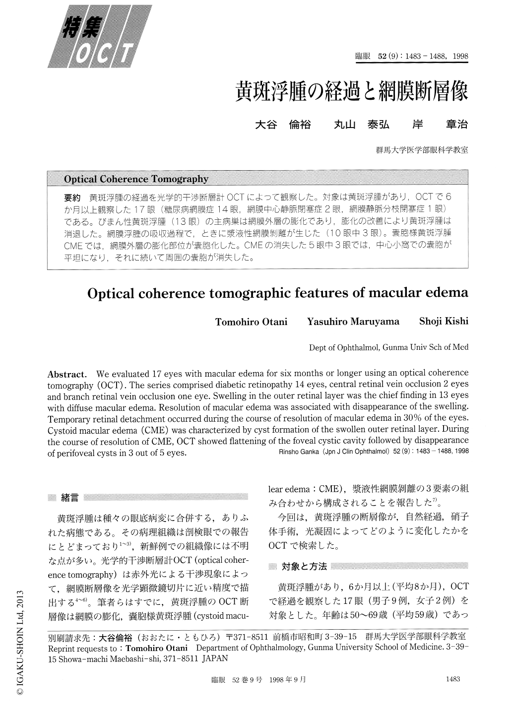
黄斑浮腫の経過を光学的干渉断層計OCTによって観察した。対象は黄斑浮腫があり,OCTで6か月以上観察した17眼(糖尿病網膜症14眼,網膜中心静脈閉塞症2眼,網膜静脈分枝閉塞症1眼)である。びまん性黄斑浮腫(13眼)の主病巣は網膜外層の膨化であり,膨化の改善により黄斑浮腫は消退した。網膜浮腫の吸収過程で,ときに漿液性網膜剥離が生じた(10眼中3眼)。嚢胞様黄斑浮腫CMEでは,網膜外層の膨化部位が嚢胞化した。CMEの消失した5眼中3眼では,中心小窩での嚢胞が平坦になり,それに続いて周囲の嚢胞が消失した。
We evaluated 17 eyes with macular edema for six months or longer using an optical coherence tomography (OCT) . The series comprised diabetic retinopathy 14 eyes, central retinal vein occlusion 2 eyes and branch retinal vein occlusion one eye. Swelling in the outer retinal layer was the chief finding in 13 eyes with diffuse macular edema. Resolution of macular edema was associated with disappearance of the swelling. Temporary retinal detachment occurred during the course of resolution of macular edema in 30% of the eyes. Cystoid macular edema (CME) was characterized by cyst formation of the swollen outer retinal layer. During the course of resolution of CME, OCT showed flattening of the foveal cystic cavity followed by disappearance of perifoveal cysts in 3 out of 5 eyes.
Copyright © 1998, Igaku-Shoin Ltd. All rights reserved.