Japanese
English
- 有料閲覧
PPV購入案内
1,320円 (1,200円+税10%) こちらは、1381ページから1384ページの文献です。購入には医書.jp本会員登録が必要です。
会員登録完了後に改めて上記「購入サイトへ」を選択してください。
または「購入サイトへ」選択後に「購入ログイン」、「新規会員登録」の順に進んでください。
会員登録について詳しくはをご確認ください。
- Abstract 文献概要
- 1ページ目 Look Inside
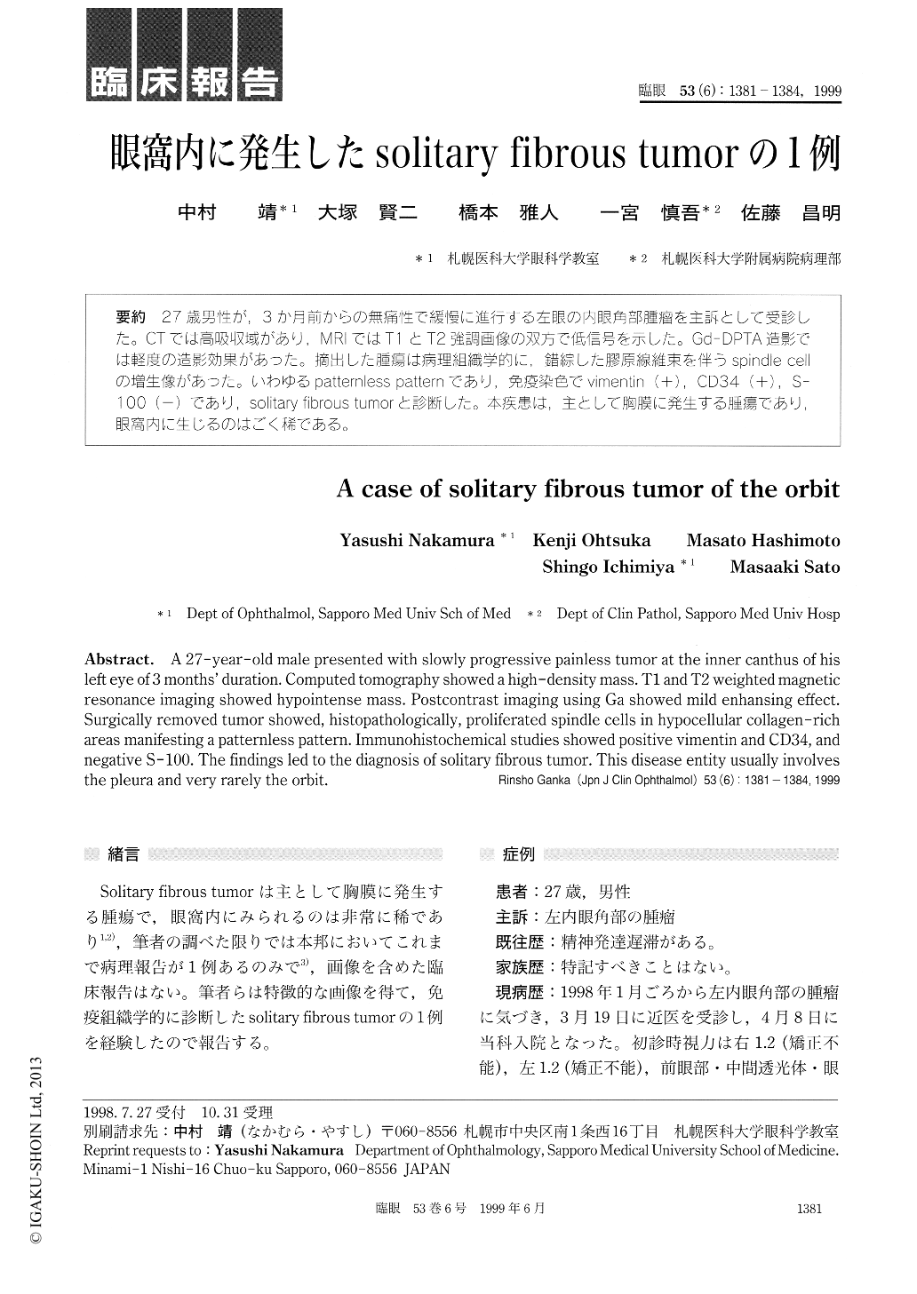
27歳男性が,3か月前からの無痛性で緩慢に進行する左眼の内眼角部腫瘤を主訴として受診した。CTでは高吸収域があり,MRIではT1とT2強調画像の双方で低信号を示した。Gd-DPTA造影では軽度の造影効果があった。摘出した腫瘍は病理組織学的に,錯綜した膠原線維束を伴うspindle cellの増生像があった。いわゆるpatternless patternであり作免疫染色でvimentin (+),CD34(+),S-100(−)であり,solitary fibrous tumorと診断した。本疾患は,主として胸膜に発生する腫瘍であり,眼窩内に生じるのはごく稀である。
A 27-year-old male presented with slowly progressive painless tumor at the inner canthus of his left eye of 3 months' duration. Computed tomography showed a high-density mass. T1 and T2 weighted magnetic resonance imaging showed hypointense mass. Postcontrast imaging using Ga showed mild enhansing effect. Surgically removed tumor showed, histopathologically, proliferated spindle cells in hypocellular collagen-rich areas manifesting a patternless pattern. Immunohistochemical studies showed positive vimentin and CD34, and negative S-100. The findings led to the diagnosis of solitary fibrous tumor. This disease entity usually involves the pleura and very rarely the orbit.
Copyright © 1999, Igaku-Shoin Ltd. All rights reserved.