Japanese
English
臨床報告
出血性眼窩リンパ管腫小児例のMRI所見
Magnetic resonance imaging of hemorrhagic orbital lymphangioma in a child
橋本 雅人
1
,
大塚 賢二
1
,
中村 靖
1
,
三浦 道子
1
,
中川 喬
1
Masato Hashimoto
1
,
Kenji Ohtsuka
1
,
Yasushi Nakamura
1
,
Michiko Miura
1
,
Takashi Nakagawa
1
1札幌医科大学医学部眼科学教室
1Dept of Ophthalmol, Sapporo Med Univ, Sch of Med
pp.1481-1484
発行日 1997年8月15日
Published Date 1997/8/15
DOI https://doi.org/10.11477/mf.1410908896
- 有料閲覧
PPV購入案内
1,320円 (1,200円+税10%) こちらは、1481ページから1484ページの文献です。
PPV(ペイ・パー・ビュー)とは、論文(記事)単位で購入・閲覧ができるサービスです。
購入には医書.jp本会員登録が必要です。
会員登録完了後に改めて上記「購入サイトへ」を選択してください。
または「購入サイトへ」選択後に「購入ログイン」、「新規会員登録」の順に進んでください。
会員登録について詳しくはをご確認ください。
購入には医書.jp本会員登録が必要です。
会員登録完了後に改めて上記「購入サイトへ」を選択してください。
または「購入サイトへ」選択後に「購入ログイン」、「新規会員登録」の順に進んでください。
会員登録について詳しくはをご確認ください。
- Abstract 文献概要
- 1ページ目 Look Inside
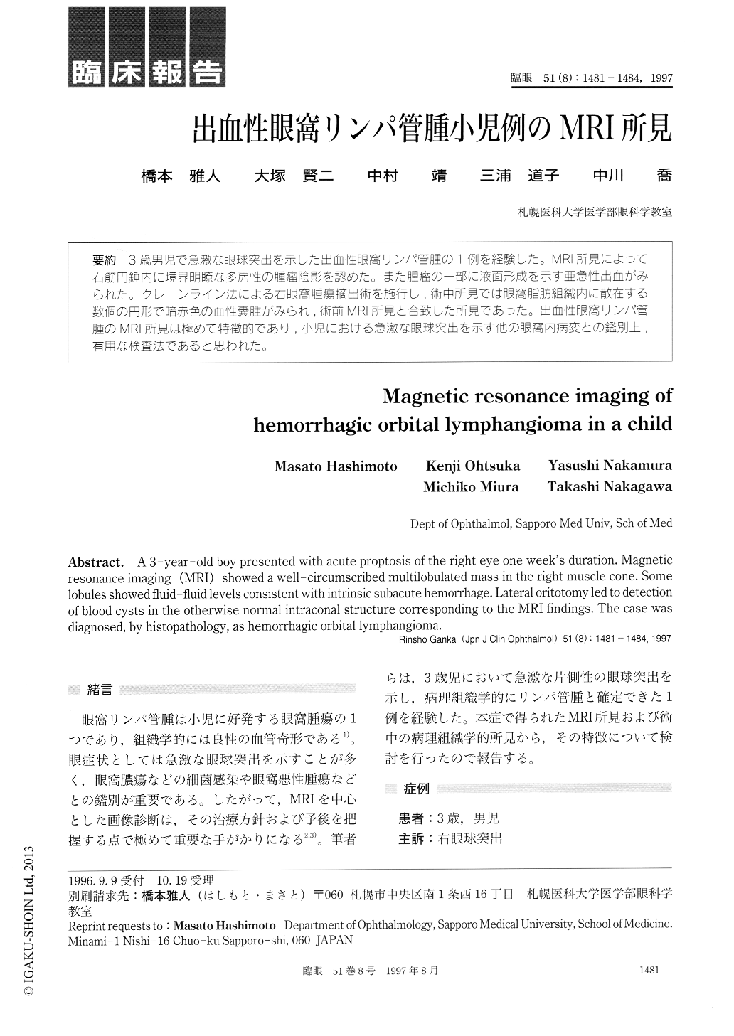
3歳男児で急激な眼球突出を示した出血性眼窩リンパ管腫の1例を経験した。MRI所見によって右筋円錘内に境界明瞭な多房性の腫瘤陰影を認めた。また腫瘤の一部に液面形成を示す亜急性出血がみられた。クレーンライン法による右眼窩腫瘍摘出術を施行し,術中所見では眼窩脂肪組織内に散在する数個の円形で暗赤色の血性嚢腫がみられ,術前MRI所見と合致した所見であった。出血性眼窩リンパ管腫のMRI所見は極めて特徴的であり,小児における急激な眼球突出を示す他の眼窩内病変との鑑別上,有用な検査法であると思われた。
A 3-year-old boy presented with acute proptosis of the right eye one week's duration. Magnetic resonance imaging (MRI) showed a well-circumscribed multilobulated mass in the right muscle cone. Some lobules showed fluid-fluid levels consistent with intrinsic subacute hemorrhage. Lateral oritotomy led to detection of blood cysts in the otherwise normal intraconal structure corresponding to the MRI findings. The case was diagnosed, by histopathology, as hemorrhagic orbital lymphangioma.
Copyright © 1997, Igaku-Shoin Ltd. All rights reserved.