Japanese
English
- 有料閲覧
PPV購入案内
1,320円 (1,200円+税10%) こちらは、1113ページから1116ページの文献です。購入には医書.jp本会員登録が必要です。
会員登録完了後に改めて上記「購入サイトへ」を選択してください。
または「購入サイトへ」選択後に「購入ログイン」、「新規会員登録」の順に進んでください。
会員登録について詳しくはをご確認ください。
- Abstract 文献概要
- 1ページ目 Look Inside
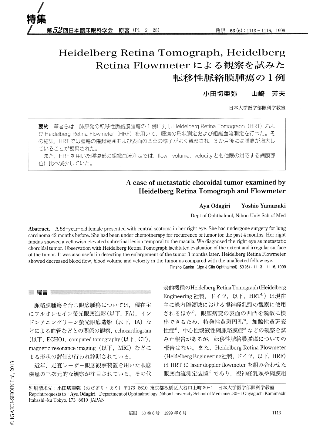
(P1-2-28) 筆者らは,肺原発の転移性脈絡膜腫瘍の1例に対しHeidelberg Retina Tomograph (HRT)およびHeidelberg Retina Flowmeter (HRF)を用いて,腫瘍の形状測定および組織血流測定を行った。その結果,HRTでは腫瘍の隆起範囲および表面の凹凸の様子がよく観察され,3か月後には腫瘍が増大していることが観察された。
また,HRFを用いた腫瘍部の組織血流測定では,flow,volume, velocityとも他眼の対応する網膜部位に比べ減少していた。
A 58-year-old female presented with central scotoma in her right eye. She had undergone surgery for lung carcinoma 42 months before. She had been under chemotherapy for recurrence of tumor for the past 4 months. Her right fundus showed a yellowish elevated subretinal lesion temporal to the macula. We diagnosed the right eye as metastatic choroidal tumor. Observation with Heidelberg Retina Tomograph facilitated evaluation of the extent and irregular surface of the tumor. It was also useful in detecting the enlargement of the tumor 3 months later. Heidelberg Retina Flowmeter showed decreased blood flow, blood volume and velocity in the tumor as compared with the unaffected fellow eye.
Copyright © 1999, Igaku-Shoin Ltd. All rights reserved.