Japanese
English
- 有料閲覧
PPV購入案内
1,320円 (1,200円+税10%) こちらは、405ページから408ページの文献です。購入には医書.jp本会員登録が必要です。
会員登録完了後に改めて上記「購入サイトへ」を選択してください。
または「購入サイトへ」選択後に「購入ログイン」、「新規会員登録」の順に進んでください。
会員登録について詳しくはをご確認ください。
- Abstract 文献概要
- 1ページ目 Look Inside
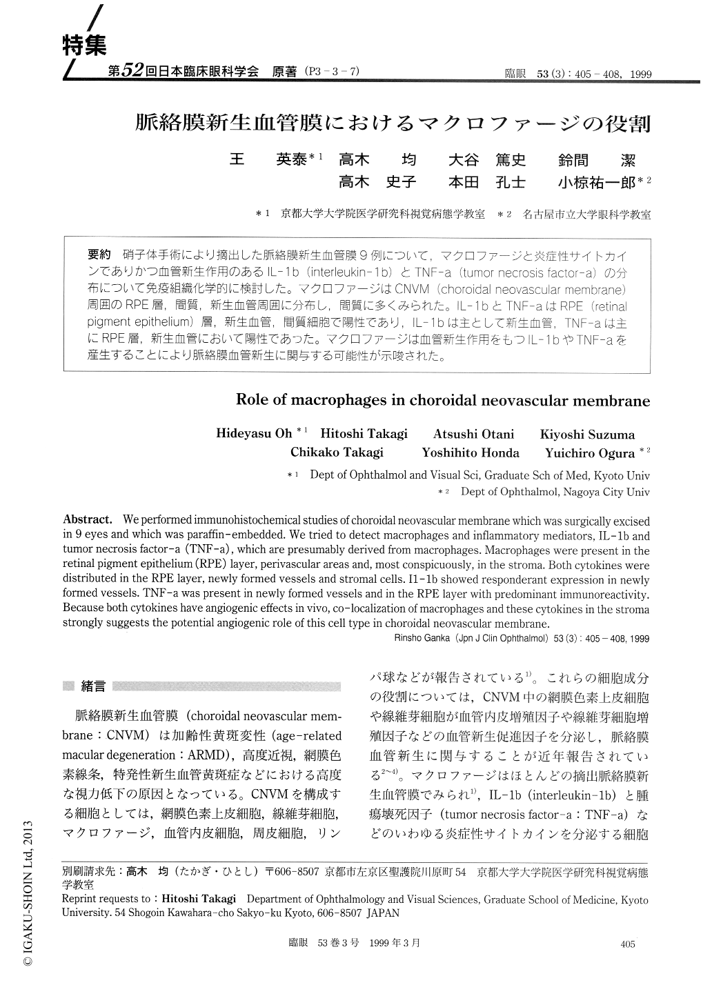
(P3-3-7) 硝子体手術により摘出した脈絡膜新生血管膜9例について,マクロファージと炎症性サイトカインでありかつ血管新生作用のあるIL-1b(interieukin-1b)とTNF-a (tumor necrosis factor-a)の分布について免疫組織化学的に検討した。マクロファージはCNVM (choroidal neovascuiar membrane)周囲のRPE層,間質,新生血管周囲に分布し,間質に多くみられた。IL-1bとTNF-aはRPE (retina-pigment epithelium)層,新生血管,間質細胞で陽性であり,IL-1bは主として新生血管,TNF-aは主にRPE層,新生血管において陽性であった。マクロファージは血管新生作用をもつIL-1bやTNF-aを産生することにより脈絡膜血管新生に関与する可能性が示唆された。
We performed immunohistochemical studies of choroidal neovascular membrane which was surgically excised in 9 eyes and which was paraffin-embedded. We tried to detect macrophages and inflammatory mediators, IL-1b and tumor necrosis factor-a (TNF-a), which are presumably derived from macrophages. Macrophages were present in the retinal pigment epithelium (RPE) layer, perivascular areas and, most conspicuously, in the stroma. Both cytokines were distributed in the RPE layer, newly formed vessels and stromal cells. I1-1b showed responderant expression in newly formed vessels. TNF-a was present in newly formed vessels and in the RPE layer with predominant immunoreactivity. Because both cytokines have angiogenic effects in vivo, co-localization of macrophages and these cytokines in the stroma strongly suggests the potential angiogenic role of this cell type in choroidal neovascular membrane.
Copyright © 1999, Igaku-Shoin Ltd. All rights reserved.