Japanese
English
- 有料閲覧
PPV購入案内
1,320円 (1,200円+税10%) こちらは、355ページから357ページの文献です。購入には医書.jp本会員登録が必要です。
会員登録完了後に改めて上記「購入サイトへ」を選択してください。
または「購入サイトへ」選択後に「購入ログイン」、「新規会員登録」の順に進んでください。
会員登録について詳しくはをご確認ください。
- Abstract 文献概要
- 1ページ目 Look Inside
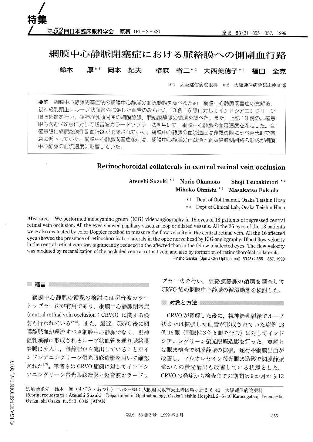
(P1-2-43) 網膜中心静脈閉塞症後の網膜中心静脈の血流動態を調べるため,網膜中心静脈閉塞症の寛解後,視神経乳頭上にループ状血管や拡張した血管のみられた13例16眼に対してインドシアニングリーン眼底造影を行い,視神経乳頭周囲の網膜静脈,脈絡膜静脈の循環を調べた。また,上記13例の非罹患眼も含む26眼に対して超音波カラードップラー法を用いて,網膜中心静脈の血流速度を測定した。全罹患眼に網脈絡膜側副血行路が形成されていた。網膜中心静脈の血流速度は非罹患眼に比べ罹患眼で有意に低下していた。網膜中心静脈閉塞症後には,網膜中心静脈の再疎通と網脈絡膜側副路の形成が網膜中心静脈の血流速度に影響していた。
We performed indocyanine green (ICG) videoangiography in 16 eyes of 13 patients of regressed central retinal vein occlusion. All the eyes showed papillary vascular loop or dilated vessels. All the 26 eyes of the 13 patients were also evaluated by color Doppler method to measure the flow velocity in the central retinal vein. All the 16 affected eyes showed the presence of retinochoroidal collaterals in the optic nerve head by ICG angiography. Blood flow velocity in the central retinal vein was significantly reduced in the affected than in the fellow unaffected eyes. The flow velocity was modified by recanalization of the occluded central retinal vein and also by formation of retinochoroidal collaterals.
Copyright © 1999, Igaku-Shoin Ltd. All rights reserved.