Japanese
English
- 有料閲覧
PPV購入案内
1,320円 (1,200円+税10%) こちらは、595ページから598ページの文献です。購入には医書.jp本会員登録が必要です。
会員登録完了後に改めて上記「購入サイトへ」を選択してください。
または「購入サイトへ」選択後に「購入ログイン」、「新規会員登録」の順に進んでください。
会員登録について詳しくはをご確認ください。
- Abstract 文献概要
- 1ページ目 Look Inside
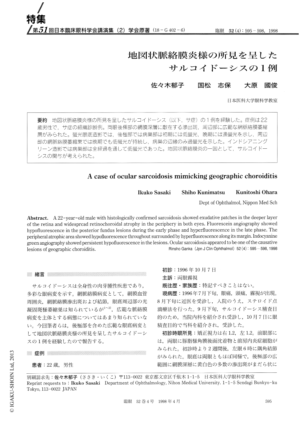
(18-G402-6) 地図状脈絡膜炎様の所見を呈したサルコイドーシス(以下,サ症)の1例を経験した。症例は22歳男性で,サ症の組織診断例。両眼後極部の網膜深層に散在する滲出斑,周辺部に広範な網脈絡膜萎縮層がみられた。螢光眼底造影では,後極部では病巣部は初期には低螢光,晩期には過螢光を示し,周辺部の網脈絡膜萎縮巣では晩期でも低螢光が持続し,病巣の辺縁のみ過螢光を示した。インドシアニングリーン造影では病巣部は全経過を通して低螢光であった。地図状脈絡膜炎の一因として,サルコイドーシスの関与が考えられた。
A 22-year-old male with histologically confirmed sarcoidosis showed exudative patches in the deeper layer of the retina and widespread retinochoroidal atrophy in the periphery in both eyes. Fluorescein angiography showed hypofluorescence in the posterior fundus lesions during the early phase and hyperfluorescence in the late phase. The peripheral atrophic area showed hypofluorescence throughout surrounded by hyperfluorescence along its margin. Indocyanine green angiography showed persistent hypofluorescence in the lesions. Ocular sarcoidosis appeared to be one of the causative lesions of geographic choroiditis.
Copyright © 1998, Igaku-Shoin Ltd. All rights reserved.