Japanese
English
- 有料閲覧
- Abstract 文献概要
- 1ページ目 Look Inside
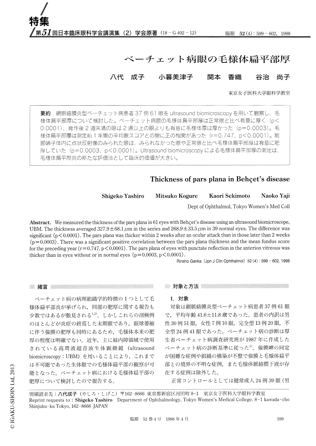
(18-G402-12) 網脈絡膜炎型ベーチェット病患者37例61眼をultrasound biomicroscopyを用いて観察し,毛様体扁平部厚について検討した。ベーチェット病眼の毛様体扁平部厚は正常眼と比べ有意に厚く(p<0.0001),発作後2週未満の眼は2週以上の眼よりも有意に毛様体厚は厚かった(p=0.0003)。毛様体扁平部厚は測定前1年間の平均眼スコアとの問に正の相関があった(r=0.747,p<0.0001)。前部硝子体内に点状反射像のみられた眼は,みられなかった眼や正常眼と比べ毛様体扁平部厚は有意に肥厚していた(p=O.OO3,p<0.0001)。Ultrasound biomicroscopyによる毛様体扁平部厚の測定は,毛様体扁平部炎の新たな評価法として臨床的価値が大きい。
We measured the thickness of the pars plana in 61 eyes with Behçet's disease using an ultrasound biomicroscope, UBM. The thickness averaged 327.9±68.1 μm in the series and 268.9 ± 33.5 gm in 39 normal eyes. The difference was significant (p < 0.0001) . The pars plana was thicker within 2 weeks after an ocular attack than in those later than 2 weeks (p = 0.0003) . There was a significant positive correlation between the pars plana thickness and the mean fundus score for the preceding year (r=0.747, p < 0.0001). The pars plana of eyes with punctate reflection in the anterion vitreous was thicker than in eyes without or in normal eyes (p=0.0003, p < 0.0001) .

Copyright © 1998, Igaku-Shoin Ltd. All rights reserved.


