Japanese
English
- 有料閲覧
PPV購入案内
1,320円 (1,200円+税10%) こちらは、55ページから61ページの文献です。購入には医書.jp本会員登録が必要です。
会員登録完了後に改めて上記「購入サイトへ」を選択してください。
または「購入サイトへ」選択後に「購入ログイン」、「新規会員登録」の順に進んでください。
会員登録について詳しくはをご確認ください。
- Abstract 文献概要
- 1ページ目 Look Inside
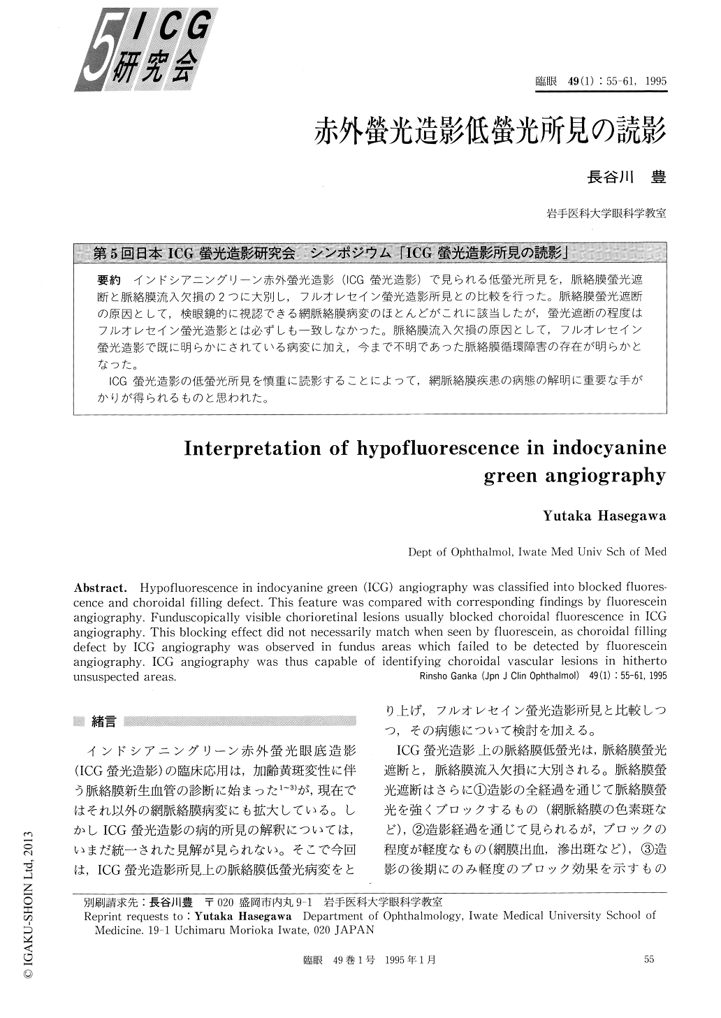
インドシアニングリーン赤外螢光造影(ICG螢光造影)で見られる低螢光所見を,脈絡膜螢光遮断と脈絡膜流入欠損の2つに大別し,フルオレセイン螢光造影所見との比較を行った。脈絡膜螢光遮断の原因として,検眼鏡的に視認できる網脈絡膜病変のほとんどがこれに該当したが,螢光遮断の程度はフルオレセイン螢光造影とは必ずしも一致しなかった。脈絡膜流入欠損の原因として,フルオレセイン螢光造影で既に明らかにされている病変に加え,今まで不明であった脈絡膜循環障害の存在が明らかとなった。
ICG螢光造影の低螢光所見を慎重に読影することによって,網脈絡膜疾患の病態の解明に重要な手がかりが得られるものと思われた。
Hypofluorescence in indocyanine green (ICG) angiography was classified into blocked fluores-cence and choroidal filling defect. This feature was compared with corresponding findings by fluorescein angiography. Funduscopically visible chorioretinal lesions usually blocked choroidal fluorescence in ICG angiography. This blocking effect did not necessarily match when seen by fluorescein, as choroidal filling defect by ICG angiography was observed in fundus areas which failed to be detected by fluorescein angiography. ICG angiography was thus capable of identifying choroidal vascular lesions in hitherto unsuspected areas.
Copyright © 1995, Igaku-Shoin Ltd. All rights reserved.