Japanese
English
- 有料閲覧
PPV購入案内
1,320円 (1,200円+税10%) こちらは、1405ページから1408ページの文献です。購入には医書.jp本会員登録が必要です。
会員登録完了後に改めて上記「購入サイトへ」を選択してください。
または「購入サイトへ」選択後に「購入ログイン」、「新規会員登録」の順に進んでください。
会員登録について詳しくはをご確認ください。
- Abstract 文献概要
- 1ページ目 Look Inside
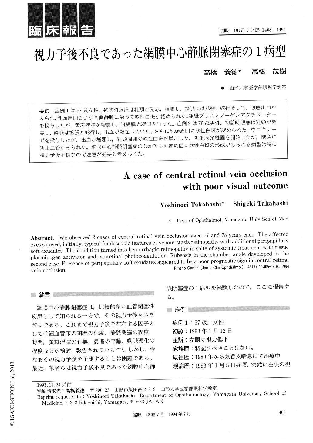
症例1は57歳女性。初診時眼底は乳頭が発赤,腫脹し,静脈には拡張,蛇行そして,眼底出血がみられ,乳頭周囲および耳側静脈に沿って軟性白斑が認められた。組織プラスミノーゲンアクチベーターを投与したが,黄斑浮腫が増悪し,汎網膜光凝固を行った。症例2は78歳男性。初診時眼底は乳頭が発赤し,静脈は拡張と蛇行し,出血が散在していた。さらに乳頭周囲に軟性白斑が認められた。ウロキナーゼを投与したが,出血が増悪し,乳頭周囲の軟性白斑が増加した。汎網膜光凝固を開始したが,隅角に新生血管がみられた。網膜中心静脈閉塞症のなかでも乳頭周囲に軟性白斑の形成がみられる病型は特に視力予後不良なので注意が必要と考えられた。
We observed 2 cases of central retinal vein occlusion aged 57 and 78 years each. The affectedeyes showed, initially, typical funduscopic features of venous stasis retinopathy with additional peripapillarysoft exudates. The condition turned into hemorrhagic retinopathy in spite of systemic treatment with tissueplasminogen activator and panretinal photocoagulation. Rubeosis in the chamber angle developed in thesecond case. Presence of peripapillary soft exudates appeared to be a poor prognostic sign in central retinalvein occlusion.
Copyright © 1994, Igaku-Shoin Ltd. All rights reserved.