Japanese
English
- 有料閲覧
PPV購入案内
1,320円 (1,200円+税10%) こちらは、297ページから306ページの文献です。購入には医書.jp本会員登録が必要です。
会員登録完了後に改めて上記「購入サイトへ」を選択してください。
または「購入サイトへ」選択後に「購入ログイン」、「新規会員登録」の順に進んでください。
会員登録について詳しくはをご確認ください。
- Abstract 文献概要
- 1ページ目 Look Inside
- 参考文献 Reference
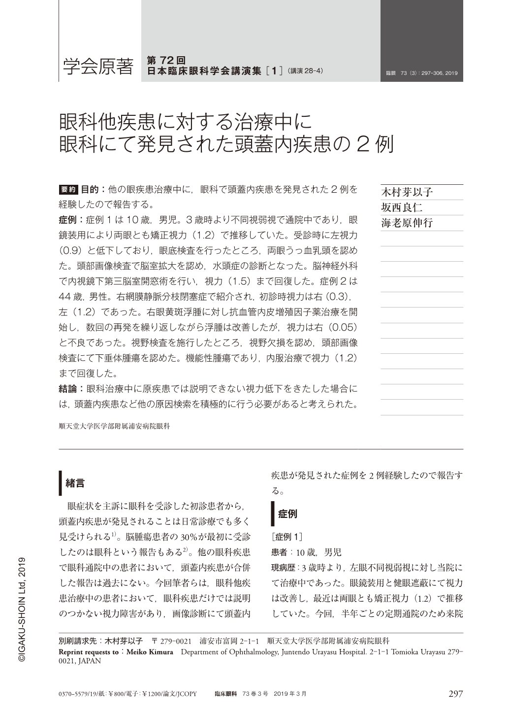
要約 目的:他の眼疾患治療中に,眼科で頭蓋内疾患を発見された2例を経験したので報告する。
症例:症例1は10歳,男児。3歳時より不同視弱視で通院中であり,眼鏡装用により両眼とも矯正視力(1.2)で推移していた。受診時に左視力(0.9)と低下しており,眼底検査を行ったところ,両眼うっ血乳頭を認めた。頭部画像検査で脳室拡大を認め,水頭症の診断となった。脳神経外科で内視鏡下第三脳室開窓術を行い,視力(1.5)まで回復した。症例2は44歳,男性。右網膜静脈分枝閉塞症で紹介され,初診時視力は右(0.3),左(1.2)であった。右眼黄斑浮腫に対し抗血管内皮増殖因子薬治療を開始し,数回の再発を繰り返しながら浮腫は改善したが,視力は右(0.05)と不良であった。視野検査を施行したところ,視野欠損を認め,頭部画像検査にて下垂体腫瘍を認めた。機能性腫瘍であり,内服治療で視力(1.2)まで回復した。
結論:眼科治療中に原疾患では説明できない視力低下をきたした場合には,頭蓋内疾患など他の原因検索を積極的に行う必要があると考えられた。
Abstract Purpose:To report two cases who were diagnosed with intracranial lesions during the course of treatment for ophthalmological conditions.
Cases and Findings:One was a 10-year-old boy. He had been treated by us for anisometropic amblyopia since the age of 3 years. Best corrected visual acuity(BCVA)improved to 1.5 in either eye after wearing eyeglasses. During follow-up, BCVA in the left eye decreased to 0.9. Funduscopy showed papilledema. Computed tomography of the head showed obstructive hydrocephalus. BCVA improved to 1.2 after endoscopic third ventriculostomy. The other was a 44-year-old male who was referred to us for branch retinal vein occlusion. BCVA was 0.3 right and 1.2 left. Intravitreal anti-vascular endothelial growth factor(VEGF)was followed by improved BCVA. BCVA in the right eye decreased to 0.05 13 months after the initial treatment and resolution of macular edema. Further examinations led to detection of pituitary tumor. Medical treatment led to improved BCVA of 1.2.
Conclusion:The present cases illustrate that intracranial lesions may be present in patients with ophthalmological diseases.
Copyright © 2019, Igaku-Shoin Ltd. All rights reserved.