Japanese
English
- 有料閲覧
PPV購入案内
1,320円 (1,200円+税10%) こちらは、91ページから101ページの文献です。購入には医書.jp本会員登録が必要です。
会員登録完了後に改めて上記「購入サイトへ」を選択してください。
または「購入サイトへ」選択後に「購入ログイン」、「新規会員登録」の順に進んでください。
会員登録について詳しくはをご確認ください。
- Abstract 文献概要
- 1ページ目 Look Inside
緒 言
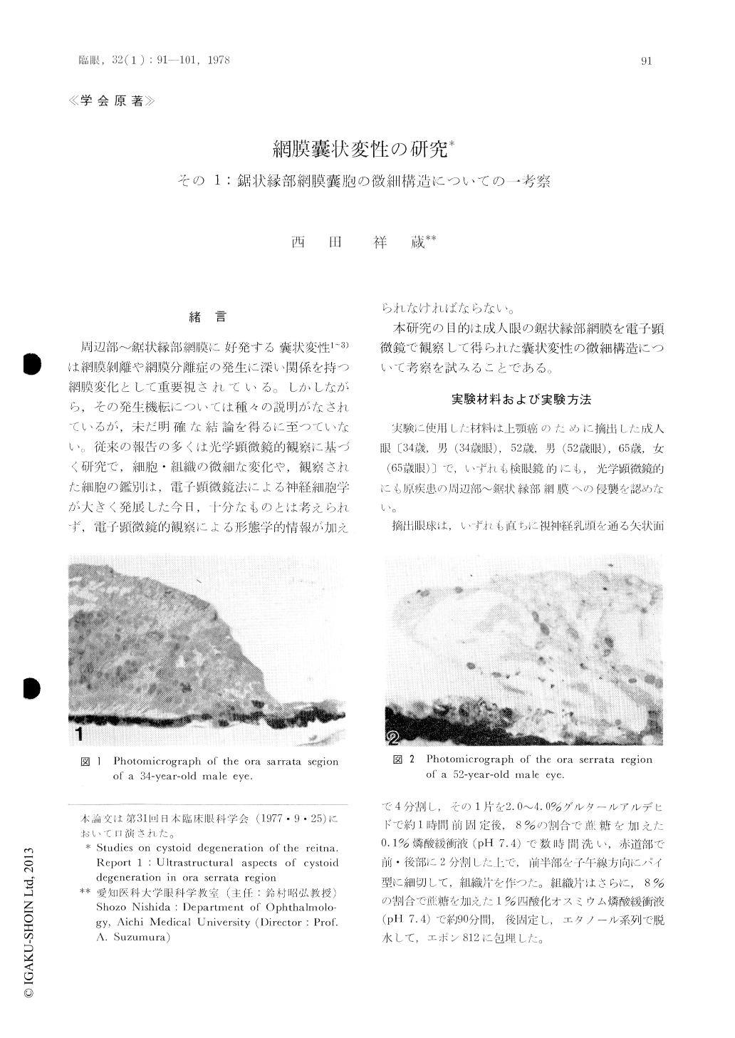
周辺部〜鋸状縁部網膜に好発する嚢状変性1〜3)は網膜剥離や網膜分離症の発生に深い関係を持つ網膜変化として重要視されている。しかしながら,その発生機転については種々の説明がなされているが,未だ明確な結論を得るに至つていない。従来の報告の多くは光学顕微鏡的観察に基づく研究で,細胞・組織の微細な変化や,観察された細胞の鑑別は,電子顕微鏡法による神経細胞学が大きく発展した今日,十分なものとは考えられず,電子顕微鏡的観察による形態学的情報が加えられなければならない。
本研究の目的は成人眼の鋸状縁部網膜を電子顕微鏡で観察して得られた嚢状変性の微細構造について考察を試みることである。
Substructural details of retinal cystoid degene-ration in the ora serrata region were studied on three adult human eyes removed due to maxillar carcinoma. Cystoid degeneration was present in the middle and inner layers of the retina. Initial cystoid degeneration seemed to occur following degenerative disappearance of neural components of the retina besides the enlargement of interspa-ces between adjacent Müller cell processes. The inner nuclear layers formed a barrier limiting the extension of the cystoid space and restricted their enlargement in the retina. Frequently, neural dendrites were exposed bare in the cystoid space.
Copyright © 1978, Igaku-Shoin Ltd. All rights reserved.