Japanese
English
- 有料閲覧
- Abstract 文献概要
- 1ページ目 Look Inside
緒言
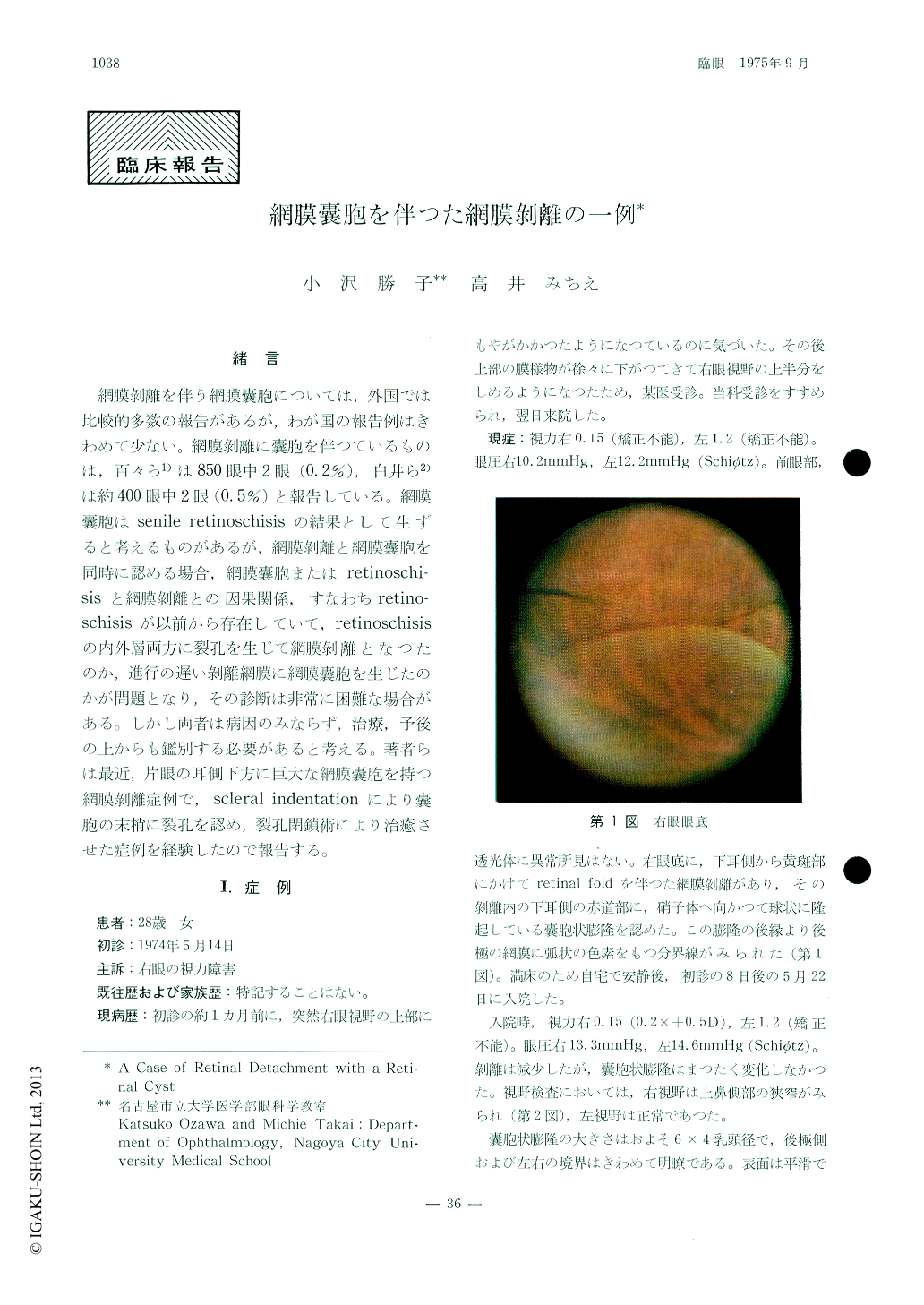
網膜剥離を伴う網膜嚢胞については,外国では比較的多数の報告があるが,わが国の報告例はきわめて少ない。網膜剥離に嚢胞を伴つているものは,百々ら1)は850眼中2眼(0.2%),白井ら2)は約400眼中2眼(0,5%)と報告している。網膜嚢胞はsenile retinoschisisの結果として生ずると考えるものがあるが,網膜剥離と網膜嚢胞を同時に認める場合,綱膜嚢胞またはretinoschi—sisと網膜剥離との因果関係,すなわちretino—schisisが以前から存在していて,retinoschisisの内外層両方に裂孔を生じて網膜剥離となつたのか,進行の遅い剥離網膜に網膜嚢胞を生じたのかが問題となり,その診断は非常に困難な場合がある。しかし両者は病因のみならず,治療,予後の上からも鑑別する必要があると考える。著者らは最近,片眼の耳側下方に巨大な網膜嚢胞を持つ網膜剥離症例で,scleral indentationにより嚢胞の末梢に裂孔を認め,裂孔閉鎖術により治癒させた症例を経験したので報告する。
This paper deals with a 28-year-old woman who had a retinal cyst difficult to differentiate from idiopathic retinoschisis. The retinal cyst was located at the lower-temporal area of the longstanding detached retina in the right eye and was 6 × 4 optic disc diameters in size. Scl-eral indentation revealed two holes at the per-ipheral fundus of the retinal cyst. There was no abnormal fluorescence at the retinal cystic region but an increase in fluorescence at the margin of retinal cyst was recognized by me-ans of fluorescence fundus angiography.

Copyright © 1975, Igaku-Shoin Ltd. All rights reserved.


