Japanese
English
- 有料閲覧
- Abstract 文献概要
- 1ページ目 Look Inside
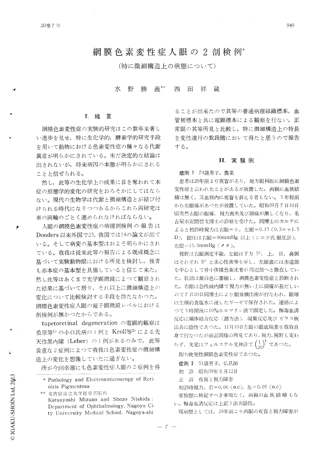
I.緒言
網膜色素変性症の実験的研究はこの数年来著しい進歩を見せ,特に生化学的,酵素学的研究手段を用いて動物における色素変性症の種々なる代謝異常が明らかにされている。未だ決定的な結論は出されないが,将来病因の本態が明らかにされることと信ぜられる。
然し,此等の生化学上の成果に目を奪われて本症の形態学的変化の研究をおろそかにしてはならない。現代の生物学は代謝と微細構造とが結び付けられる時代になりつつあるからこれら両研究は車の両輪のごとく進められなければならない。
Retinal vascular tree in a relatively advan-ced case of retinitis pigmentosa reveals ace-llularity and hyalinization of the capillary wall. Some of capillary branches show aneu-rysm-like appearance, and pigment granules lodge compactly around them.
Electronmicroscopy of the retina in this ca-se reveals that the cone outer segment has undergone every degree of degeneration, that cystic pigment proliferation has developed which contain cone remnants, fuscin granules,mitochondrion, membrane ststem. ribosomes and lysozomes. The inner segments are short and have fewer mitochondria than normal.

Copyright © 1966, Igaku-Shoin Ltd. All rights reserved.


