Japanese
English
臨床実験
細隙灯顕微鏡による眼底撮影,特に倍率について
Magnification Factors in Slitlamp Fundus Photography
三国 政吉
1
,
八百枝 浩
1
Masakichi Mikuni
1
,
Hiroshi Yaoeda
1
1新潟大学医学部眼科学教室
1Department of Ophthalmology, Niigata University School of Medicine
pp.1163-1166
発行日 1970年9月15日
Published Date 1970/9/15
DOI https://doi.org/10.11477/mf.1410204371
- 有料閲覧
PPV購入案内
1,320円 (1,200円+税10%) こちらは、1163ページから1166ページの文献です。
PPV(ペイ・パー・ビュー)とは、論文(記事)単位で購入・閲覧ができるサービスです。
購入には医書.jp本会員登録が必要です。
会員登録完了後に改めて上記「購入サイトへ」を選択してください。
または「購入サイトへ」選択後に「購入ログイン」、「新規会員登録」の順に進んでください。
会員登録について詳しくはをご確認ください。
購入には医書.jp本会員登録が必要です。
会員登録完了後に改めて上記「購入サイトへ」を選択してください。
または「購入サイトへ」選択後に「購入ログイン」、「新規会員登録」の順に進んでください。
会員登録について詳しくはをご確認ください。
- Abstract 文献概要
- 1ページ目 Look Inside
I.緒言
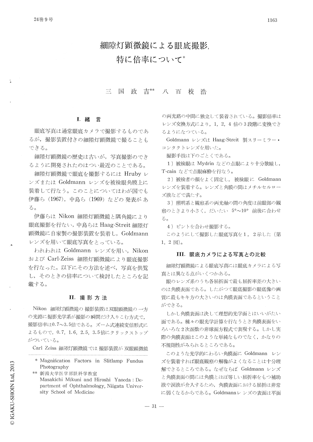
眼底写真は通常眼底カメラで撮影するものであるが,撮影装置付きの細隙灯顕微鏡で撮ることもできる。
細隙灯顕微鏡の歴史は古いが,写真撮影のできるように開発されたのはつい最近のことである。
Fundus photographs were taken by slitlamp microscopes (Nikon or Carl Zeiss) with the use of flat-faced portion of the Goldmann contact lens.
1) The theoretical magnification of the fundus picture was calculated in the optical system composed of Gullstrand's simplified schematic eye and the contact lens by means of raytrac-ing method. The estimated value was 1.075x.
2) Among factors which might influence the magnification, the refractive indices of the cry-stallin lens, the vitreous and the aqueous seem-ed to be major ones.
Copyright © 1970, Igaku-Shoin Ltd. All rights reserved.