Japanese
English
- 有料閲覧
PPV購入案内
1,320円 (1,200円+税10%) こちらは、645ページから650ページの文献です。購入には医書.jp本会員登録が必要です。
会員登録完了後に改めて上記「購入サイトへ」を選択してください。
または「購入サイトへ」選択後に「購入ログイン」、「新規会員登録」の順に進んでください。
会員登録について詳しくはをご確認ください。
- Abstract 文献概要
- 1ページ目 Look Inside
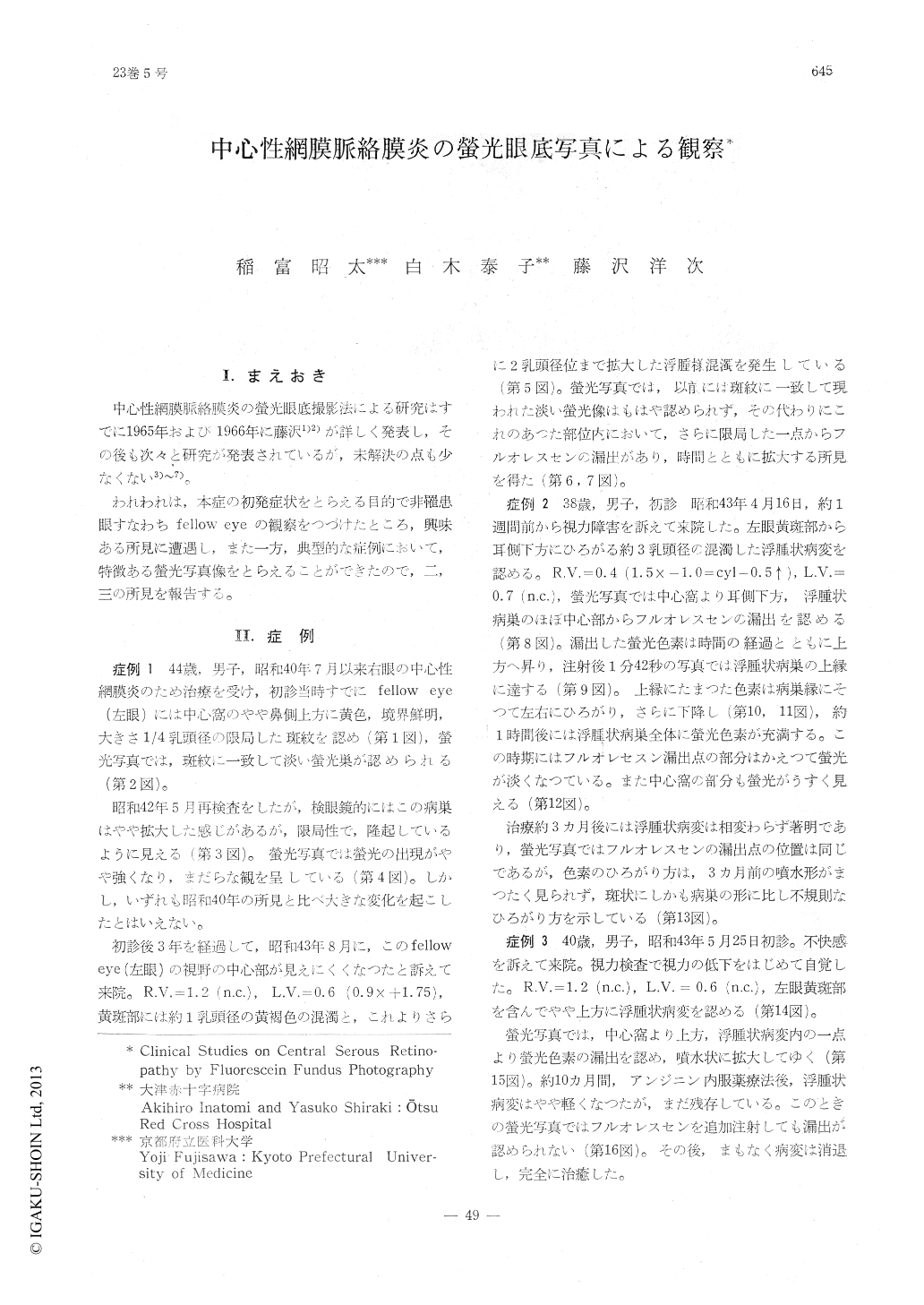
I.まえおき
中心性網膜脈絡膜炎の螢光眼底撮影法による研究はすでに1965年および1966年に藤沢1)2)が詳しく発表し,その後も次々と研究が発表されているが,未解決の点も少なくない3)〜7)。
われわれは,本症の初発症状をとらえる目的で非罹患眼すなわちfellow eyeの観察をつづけたところ,興味ある所見に遭遇し,また一方,典型的な症例において,特徴ある螢光写真像をとらえることができたので,二,三の所見を報告する。
1 ) In a case of central serous retinopathy (Masuda's disease), a focal yellow lesion of the fundus, which had been found ophthalmo-scopically in the fellow eye, developed into the typical retinal edema (more accurately, deta-chment).
The fluorescein findings of the yellow lesion suggested the presence of a detachment of the pigment epithelium, which disappeared with the abnormal increasing in its permeability and developed into the retinal detachment.
It is more likely that the detachment of the pigment epithelium is a primary change and the retinal detachment is a secondary one in central serous retinopathy.
Copyright © 1969, Igaku-Shoin Ltd. All rights reserved.