Japanese
English
臨床実験
位相差顕微鏡による硝子体所見
The Vitreous Structure. A Phase-Contrast Microscopic Study
渡部 通英
1
Michihide Watanabe
1
1東京慈恵会医科大学眼科学教室
1Department of Ophthalmology, Jikei Medical College
pp.1167-1176
発行日 1968年9月15日
Published Date 1968/9/15
DOI https://doi.org/10.11477/mf.1410203929
- 有料閲覧
PPV購入案内
1,320円 (1,200円+税10%) こちらは、1167ページから1176ページの文献です。
PPV(ペイ・パー・ビュー)とは、論文(記事)単位で購入・閲覧ができるサービスです。
購入には医書.jp本会員登録が必要です。
会員登録完了後に改めて上記「購入サイトへ」を選択してください。
または「購入サイトへ」選択後に「購入ログイン」、「新規会員登録」の順に進んでください。
会員登録について詳しくはをご確認ください。
購入には医書.jp本会員登録が必要です。
会員登録完了後に改めて上記「購入サイトへ」を選択してください。
または「購入サイトへ」選択後に「購入ログイン」、「新規会員登録」の順に進んでください。
会員登録について詳しくはをご確認ください。
- Abstract 文献概要
- 1ページ目 Look Inside
I.はじめに
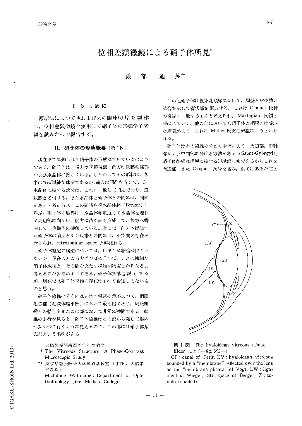
凍結法によつて豚および人の眼球切片を製作し,位相差顕微鏡を使用して硝子体の形態学的考察を試みたので報告する。
The vitreous body of human and the pig was subjected to phase-contrast-microscopic studies in frozen-cut sections without undergoing fixa-tive or staining procedures. The method enabled easy identification of the anterior limiting mem-brane of the vitreous and the canal of Cloquet. Four groups of vitreous fiber originating from the pars plana of the ciliary body could be identified.
Copyright © 1968, Igaku-Shoin Ltd. All rights reserved.