Japanese
English
- 有料閲覧
PPV購入案内
1,320円 (1,200円+税10%) こちらは、1357ページから1360ページの文献です。購入には医書.jp本会員登録が必要です。
会員登録完了後に改めて上記「購入サイトへ」を選択してください。
または「購入サイトへ」選択後に「購入ログイン」、「新規会員登録」の順に進んでください。
会員登録について詳しくはをご確認ください。
- Abstract 文献概要
- 1ページ目 Look Inside
- 参考文献 Reference
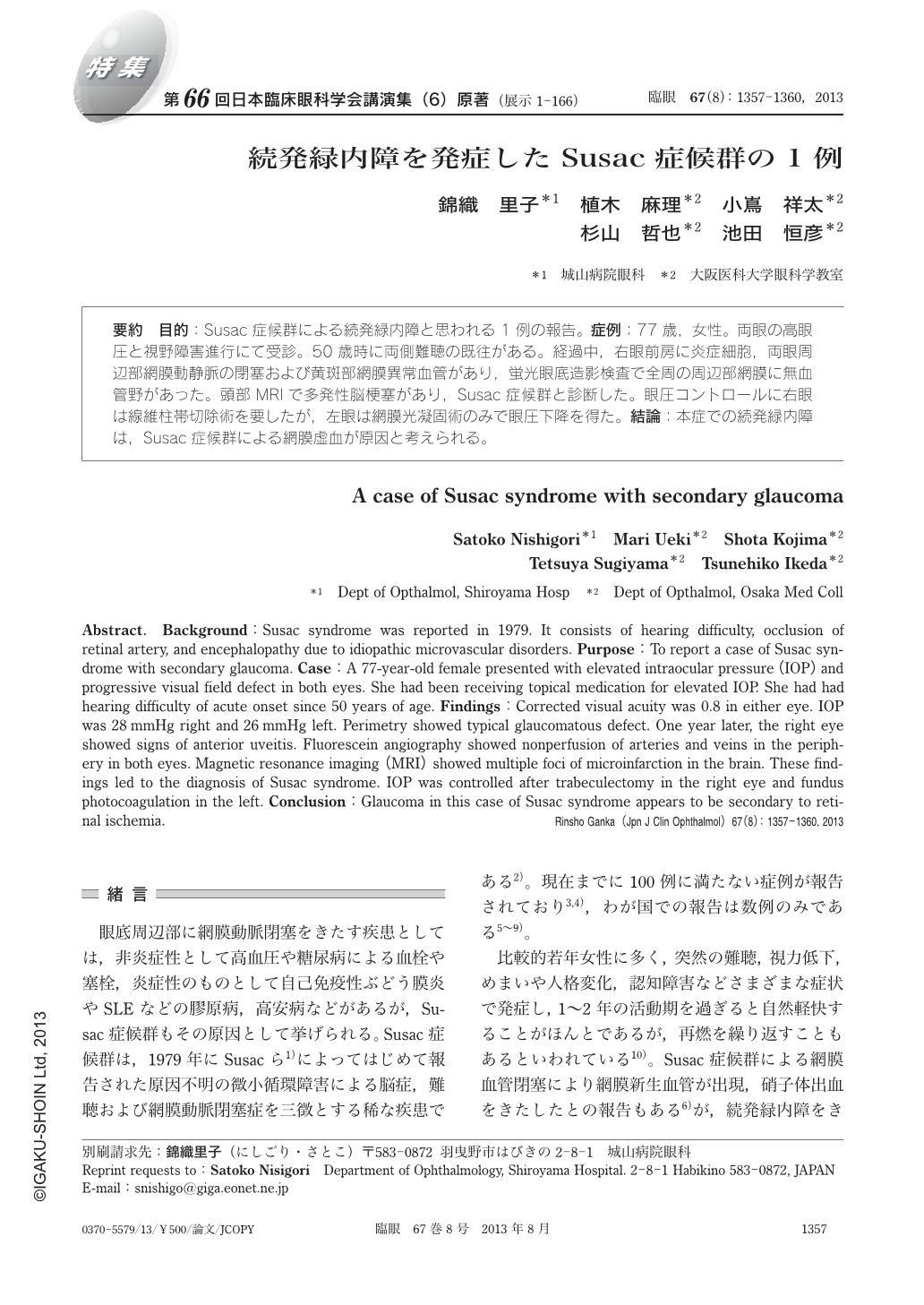
要約 目的:Susac症候群による続発緑内障と思われる1例の報告。症例:77歳,女性。両眼の高眼圧と視野障害進行にて受診。50歳時に両側難聴の既往がある。経過中,右眼前房に炎症細胞,両眼周辺部網膜動静脈の閉塞および黄斑部網膜異常血管があり,蛍光眼底造影検査で全周の周辺部網膜に無血管野があった。頭部MRIで多発性脳梗塞があり,Susac症候群と診断した。眼圧コントロールに右眼は線維柱帯切除術を要したが,左眼は網膜光凝固術のみで眼圧下降を得た。結論:本症での続発緑内障は,Susac症候群による網膜虚血が原因と考えられる。
Abstract. Background:Susac syndrome was reported in 1979. It consists of hearing difficulty, occlusion of retinal artery, and encephalopathy due to idiopathic microvascular disorders. Purpose:To report a case of Susac syndrome with secondary glaucoma. Case:A 77-year-old female presented with elevated intraocular pressure(IOP)and progressive visual field defect in both eyes. She had been receiving topical medication for elevated IOP. She had had hearing difficulty of acute onset since 50 years of age. Findings:Corrected visual acuity was 0.8 in either eye. IOP was 28 mmHg right and 26 mmHg left. Perimetry showed typical glaucomatous defect. One year later, the right eye showed signs of anterior uveitis. Fluorescein angiography showed nonperfusion of arteries and veins in the periphery in both eyes. Magnetic resonance imaging(MRI)showed multiple foci of microinfarction in the brain. These findings led to the diagnosis of Susac syndrome. IOP was controlled after trabeculectomy in the right eye and fundus photocoagulation in the left. Conclusion:Glaucoma in this case of Susac syndrome appears to be secondary to retinal ischemia.
Copyright © 2013, Igaku-Shoin Ltd. All rights reserved.