Japanese
English
- 有料閲覧
PPV購入案内
1,320円 (1,200円+税10%) こちらは、1963ページから1966ページの文献です。購入には医書.jp本会員登録が必要です。
会員登録完了後に改めて上記「購入サイトへ」を選択してください。
または「購入サイトへ」選択後に「購入ログイン」、「新規会員登録」の順に進んでください。
会員登録について詳しくはをご確認ください。
- Abstract 文献概要
- 1ページ目 Look Inside
- 参考文献 Reference
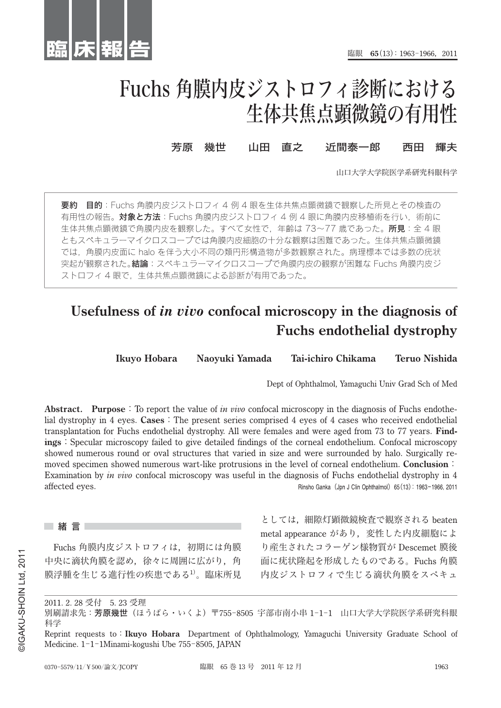
要約 目的:Fuchs角膜内皮ジストロフィ4例4眼を生体共焦点顕微鏡で観察した所見とその検査の有用性の報告。対象と方法:Fuchs角膜内皮ジストロフィ4例4眼に角膜内皮移植術を行い,術前に生体共焦点顕微鏡で角膜内皮を観察した。すべて女性で,年齢は73~77歳であった。所見:全4眼ともスペキュラーマイクロスコープでは角膜内皮細胞の十分な観察は困難であった。生体共焦点顕微鏡では,角膜内皮面にhaloを伴う大小不同の類円形構造物が多数観察された。病理標本では多数の疣状突起が観察された。結論:スペキュラーマイクロスコープで角膜内皮の観察が困難なFuchs角膜内皮ジストロフィ4眼で,生体共焦点顕微鏡による診断が有用であった。
Abstract. Purpose:To report the value of in vivo confocal microscopy in the diagnosis of Fuchs endothelial dystrophy in 4 eyes. Cases:The present series comprised 4 eyes of 4 cases who received endothelial transplantation for Fuchs endothelial dystrophy. All were females and were aged from 73 to 77 years. Findings:Specular microscopy failed to give detailed findings of the corneal endothelium. Confocal microscopy showed numerous round or oval structures that varied in size and were surrounded by halo. Surgically removed specimen showed numerous wart-like protrusions in the level of corneal endothelium. Conclusion:Examination by in vivo confocal microscopy was useful in the diagnosis of Fuchs endothelial dystrophy in 4 affected eyes.
Copyright © 2011, Igaku-Shoin Ltd. All rights reserved.