Japanese
English
- 有料閲覧
PPV購入案内
1,320円 (1,200円+税10%) こちらは、483ページから490ページの文献です。購入には医書.jp本会員登録が必要です。
会員登録完了後に改めて上記「購入サイトへ」を選択してください。
または「購入サイトへ」選択後に「購入ログイン」、「新規会員登録」の順に進んでください。
会員登録について詳しくはをご確認ください。
- Abstract 文献概要
- 1ページ目 Look Inside
- 参考文献 Reference
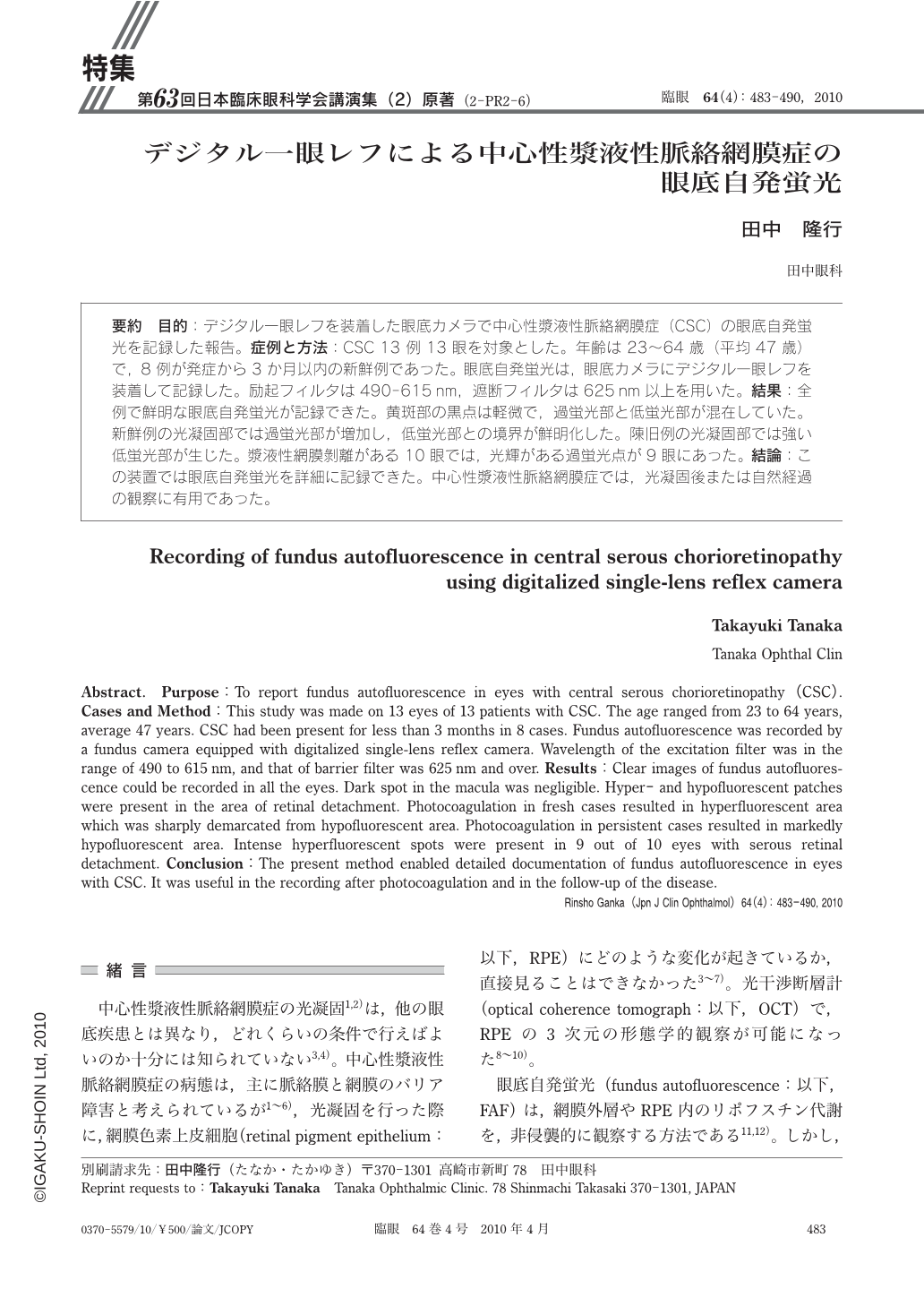
要約 目的:デジタル一眼レフを装着した眼底カメラで中心性漿液性脈絡網膜症(CSC)の眼底自発蛍光を記録した報告。症例と方法:CSC 13例13眼を対象とした。年齢は23~64歳(平均47歳)で,8例が発症から3か月以内の新鮮例であった。眼底自発蛍光は,眼底カメラにデジタル一眼レフを装着して記録した。励起フィルタは490-615nm,遮断フィルタは625nm以上を用いた。結果:全例で鮮明な眼底自発蛍光が記録できた。黄斑部の黒点は軽微で,過蛍光部と低蛍光部が混在していた。新鮮例の光凝固部では過蛍光部が増加し,低蛍光部との境界が鮮明化した。陳旧例の光凝固部では強い低蛍光部が生じた。漿液性網膜剝離がある10眼では,光輝がある過蛍光点が9眼にあった。結論:この装置では眼底自発蛍光を詳細に記録できた。中心性漿液性脈絡網膜症では,光凝固後または自然経過の観察に有用であった。
Abstract. Purpose:To report fundus autofluorescence in eyes with central serous chorioretinopathy(CSC). Cases and Method:This study was made on 13 eyes of 13 patients with CSC. The age ranged from 23 to 64 years,average 47 years. CSC had been present for less than 3 months in 8 cases. Fundus autofluorescence was recorded by a fundus camera equipped with digitalized single-lens reflex camera. Wavelength of the excitation filter was in the range of 490 to 615 nm,and that of barrier filter was 625 nm and over. Results:Clear images of fundus autofluorescence could be recorded in all the eyes. Dark spot in the macula was negligible. Hyper-and hypofluorescent patches were present in the area of retinal detachment. Photocoagulation in fresh cases resulted in hyperfluorescent area which was sharply demarcated from hypofluorescent area. Photocoagulation in persistent cases resulted in markedly hypofluorescent area. Intense hyperfluorescent spots were present in 9 out of 10 eyes with serous retinal detachment. Conclusion:The present method enabled detailed documentation of fundus autofluorescence in eyes with CSC. It was useful in the recording after photocoagulation and in the follow-up of the disease.
Copyright © 2010, Igaku-Shoin Ltd. All rights reserved.