Japanese
English
- 有料閲覧
PPV購入案内
1,320円 (1,200円+税10%) こちらは、531ページから534ページの文献です。購入には医書.jp本会員登録が必要です。
会員登録完了後に改めて上記「購入サイトへ」を選択してください。
または「購入サイトへ」選択後に「購入ログイン」、「新規会員登録」の順に進んでください。
会員登録について詳しくはをご確認ください。
- Abstract 文献概要
- 1ページ目 Look Inside
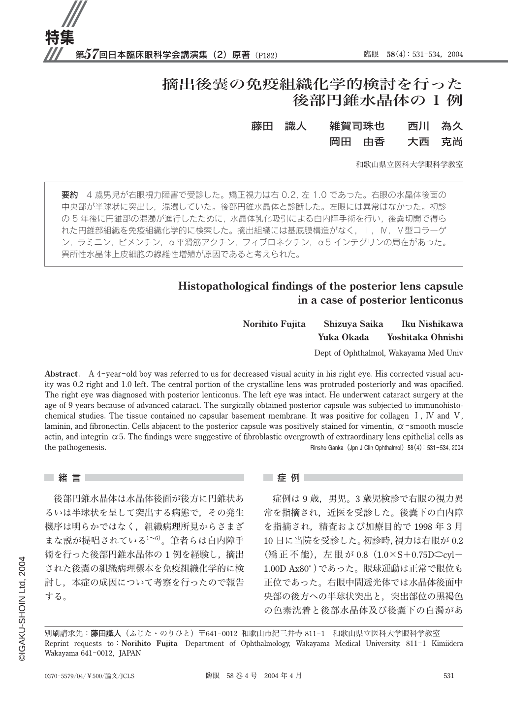
4歳男児が右眼視力障害で受診した。矯正視力は右0.2,左1.0であった。右眼の水晶体後面の中央部が半球状に突出し,混濁していた。後部円錐水晶体と診断した。左眼には異常はなかった。初診の5年後に円錐部の混濁が進行したために,水晶体乳化吸引による白内障手術を行い,後囊切開で得られた円錐部組織を免疫組織化学的に検索した。摘出組織には基底膜構造がなく,Ⅰ,Ⅳ,Ⅴ型コラーゲン,ラミニン,ビメンチン,α平滑筋アクチン,フィブロネクチン,α5インテグリンの局在があった。異所性水晶体上皮細胞の線維性増殖が原因であると考えられた。
A 4-year-old boy was referred to us for decreased visual acuity in his right eye. His corrected visual acuity was 0.2 right and 1.0 left. The central portion of the crystalline lens was protruded posteriorly and was opacified. The right eye was diagnosed with posterior lenticonus. The left eye was intact. He underwent cataract surgery at the age of 9 years because of advanced cataract. The surgically obtained posterior capsule was subjected to immunohistochemical studies. The tissue contained no capsular basement membrane. It was positive for collagenⅠ,ⅣandⅤ,laminin,and fibronectin. Cells abjacent to the posterior capsule was positively stained for vimentin,α-smooth muscle actin,and integrinα5. The findings were suggestive of fibroblastic overgrowth of extraordinary lens epithelial cells as the pathogenesis.
Copyright © 2004, Igaku-Shoin Ltd. All rights reserved.