Japanese
English
- 有料閲覧
PPV購入案内
1,320円 (1,200円+税10%) こちらは、987ページから990ページの文献です。購入には医書.jp本会員登録が必要です。
会員登録完了後に改めて上記「購入サイトへ」を選択してください。
または「購入サイトへ」選択後に「購入ログイン」、「新規会員登録」の順に進んでください。
会員登録について詳しくはをご確認ください。
- Abstract 文献概要
- 1ページ目 Look Inside
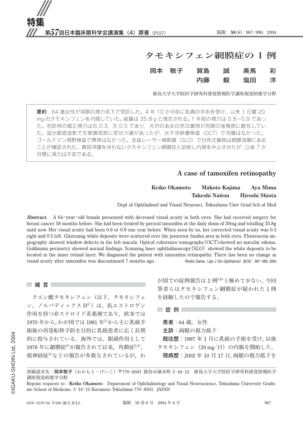
64歳女性が両眼の視力低下で受診した。4年10か月前に乳癌の手術を受け,以来1日量20mgのタモキシフェンを内服していた。総量は35.8gと推定される。1年前の視力は0.8~0.9であった。初診時の矯正視力は右0.3,左0.5であり,光沢のある白色沈着物が両眼の後極部に散在していた。蛍光眼底造影で左眼黄斑部に窓状欠損があったが,光干渉断層検査(OCT)で浮腫はなかった。ゴールドマン視野検査で異常はなかった。走査レーザー検眼鏡(SLO)で白色沈着物は網膜浅層にあることが確認された。黄斑浮腫を伴わないタモキシフェン網膜症と診断し内服を中止させたが,以後7か月間に視力は不変である。
A 64-year-old female presented with decreaed visual acuity in both eyes. She had received surgery for breast cancer 58months before. She had been treated by peroral tamoxifen at the daily dosis of 20mg and totalling 35.8g until now. Her visual acuity had been 0.8 or 0.9 one year before. When seen by us,her corrected visual acuity was 0.3 right and 0.5 left. Glistening white deposits were scattered over the posterior fundus area in both eyes. Fluorescein angiography showed window defects in the left macula. Optical coherence tomography(OCT)showed no macular edema. Goldmann perimetry showed normal findings. Scanning laser ophthalmoscopy(SLO)showed the white deposits to be located in the inner retinal layer. We diagnosed the patient with tamoxifen retinopathy. There has been no change in visual acuity after tamoxifen was discontinued 7months ago.
Copyright © 2004, Igaku-Shoin Ltd. All rights reserved.