Japanese
English
- 有料閲覧
PPV購入案内
1,320円 (1,200円+税10%) こちらは、68ページから70ページの文献です。購入には医書.jp本会員登録が必要です。
会員登録完了後に改めて上記「購入サイトへ」を選択してください。
または「購入サイトへ」選択後に「購入ログイン」、「新規会員登録」の順に進んでください。
会員登録について詳しくはをご確認ください。
- Abstract 文献概要
- 1ページ目 Look Inside
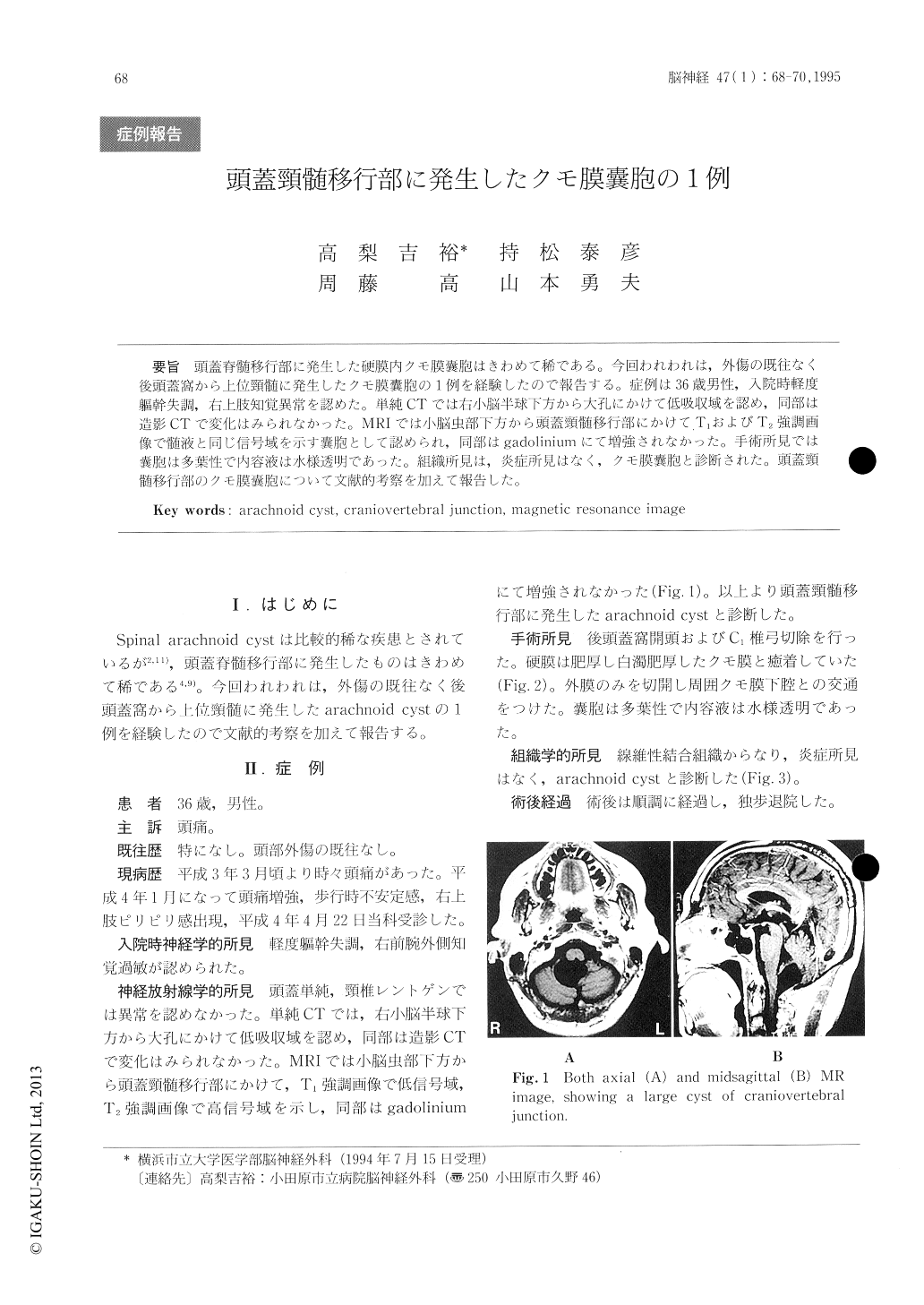
頭蓋脊髄移行部に発生した硬膜内クモ膜嚢胞はきわめて稀である。今回われわれは,外傷の既往なく後頭蓋窩から上位頸髄に発生したクモ膜嚢胞の1例を経験したので報告する。症例は36歳男性,入院時軽度躯幹失調,右上肢知覚異常を認めた。単純CTでは右小脳半球下方から大孔にかけて低吸収域を認め,同部は造影CTで変化はみられなかった。MRIでは小脳虫部下方から頭蓋頸髄移行部にかけてT1およびT2強調画像で髄液と同じ信号域を示す嚢胞として認められ,同部はgadoliniumにて増強されなかった。手術所見では嚢胞は多葉性で内容液は水様透明であった。組織所見は,炎症所見はなく,クモ膜嚢胞と診断された。頭蓋頸髄移行部のクモ膜嚢胞について文献的考察を加えて報告した。
We reported a case of arachnoid cyst in the craniovertebral junction which was extremely rare. A 36-year-old man presented truncal ataxia and dysesthesia in the right upper extremity. CT and MR images revealed a large cyst in the cranioverte-bral junction. As for findings of MR images, cystic lesion showed similar intensity as cerebrospinal fluid. Intradural arachnoid cyst with thickened dura was opened to communicate with subarachnoid space. Fluid in the cyst was waterly clear. His-tological finding of the surgical specimen was arach-noid cyst without inflammatory changes.
Copyright © 1995, Igaku-Shoin Ltd. All rights reserved.