Japanese
English
- 有料閲覧
PPV購入案内
1,320円 (1,200円+税10%) こちらは、1097ページから1099ページの文献です。購入には医書.jp本会員登録が必要です。
会員登録完了後に改めて上記「購入サイトへ」を選択してください。
または「購入サイトへ」選択後に「購入ログイン」、「新規会員登録」の順に進んでください。
会員登録について詳しくはをご確認ください。
- Abstract 文献概要
- 1ページ目 Look Inside
I.はじめに
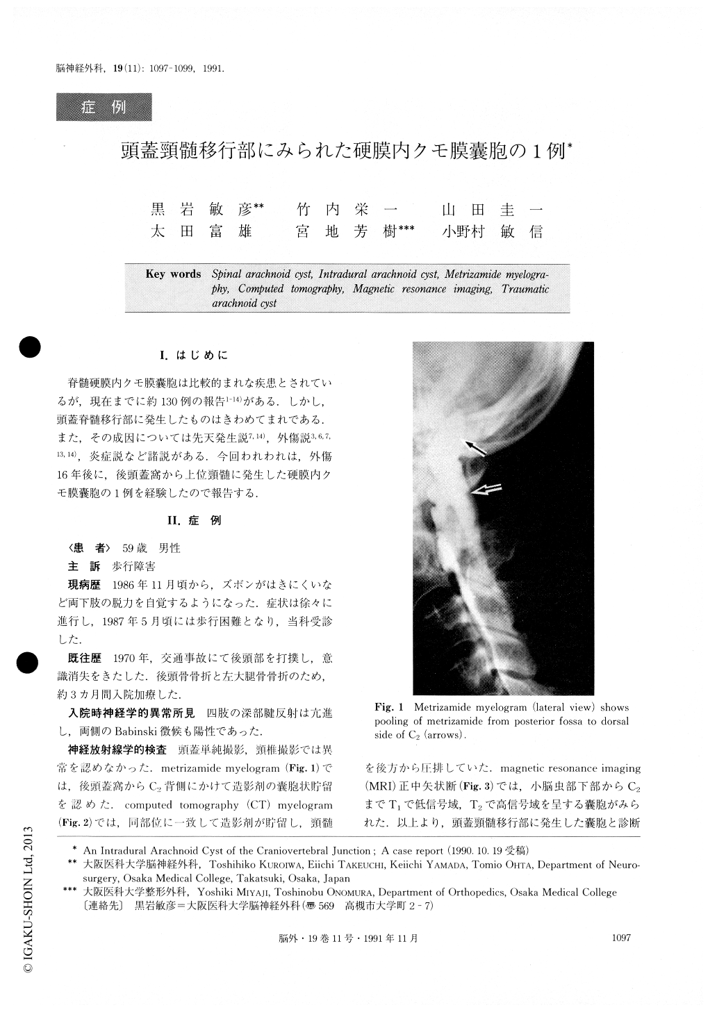
脊髄硬膜内クモ膜嚢胞は比較的まれな疾患とされているが,現在までに約130例の報告1-14)がある.しかし,頭蓋脊髄移行部に発生したものはきわめてまれである.また,その成因については先天発生説7,14)外傷説3,6,7,13,14),炎症説など諸説がある.今回われわれは,外傷16年後に,後頭蓋窩から上位頸髄に発生した硬膜内クモ膜嚢胞の1例を経験したので報告する.
Abstract
An intradural arachnoid cyst of the craniovertebral junction possibly of traumatic origin is reported. A 59-year-old man was admitted to our hospital with a 10-month history of progressive gait disturbance. He had a history of head injury with a fracture of the occipital bone. Myelography revealed pooling of the contrast medium in the posterior fossa and on the dorsal sides of C1 and C2. Metrizamide-enhanced computed tomography also showed pooling at the same level.Magnetic resonance imaging indicated a large cystic lesion at the craniovertebral junction. Craniectomy of the posterior fossa and laminectomy of C1, C2 and C3 were performed, and an intradural cyst with thickened dura and arachnoid was found. The cyst wall was opened to communicate with the subarachnoid space. Histological findings of the specimen showed that the arachnoid was thickened.
There are over 130 reports of intradural arachnoid cyst of the spine, but those of traumatic origin are rare, and cysts located in the intracranial to spinal region are extremely rare.
Copyright © 1991, Igaku-Shoin Ltd. All rights reserved.