Japanese
English
- 有料閲覧
PPV購入案内
1,320円 (1,200円+税10%) こちらは、1051ページから1057ページの文献です。購入には医書.jp本会員登録が必要です。
会員登録完了後に改めて上記「購入サイトへ」を選択してください。
または「購入サイトへ」選択後に「購入ログイン」、「新規会員登録」の順に進んでください。
会員登録について詳しくはをご確認ください。
- Abstract 文献概要
- 1ページ目 Look Inside
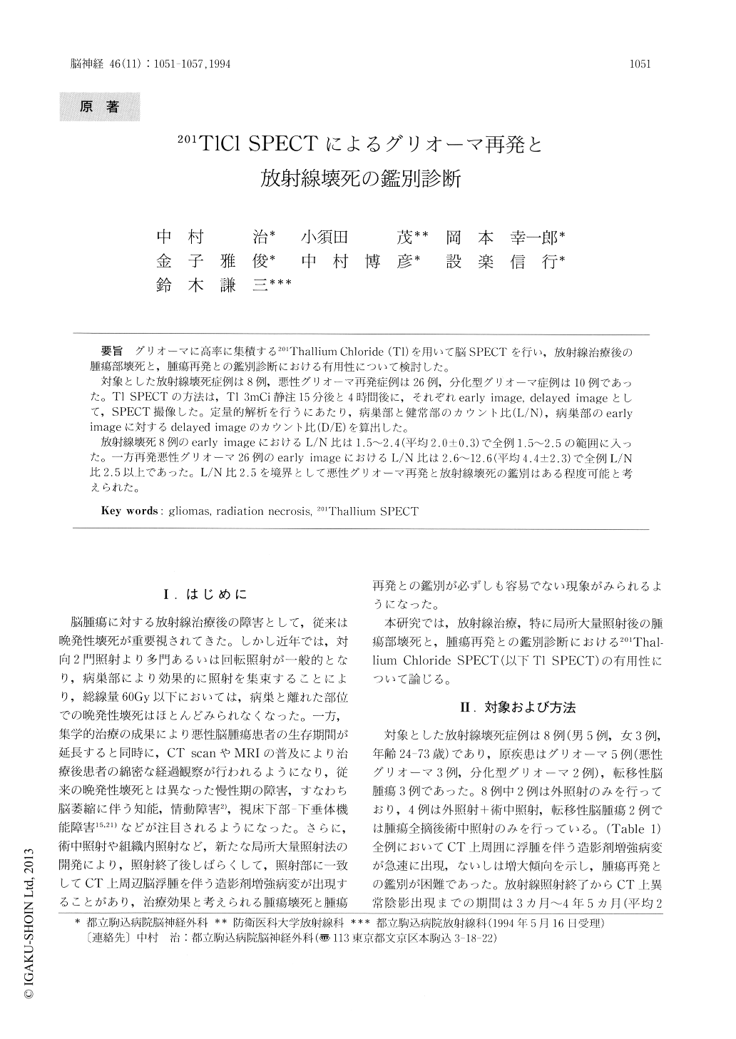
グリオーマに高率に集積する201Thallium Chloride(Tl)を用いて脳SPECTを行い,放射線治療後の腫瘍部壊死と,腫瘍再発との鑑別診断における有用性について検討した。
対象とした放射線壊死症例は8例,悪性グリオーマ再発症例は26例,分化型グリオーマ症例は10例であった。Tl SPECTの方法は,Tl 3mCi静注15分後と4時間後に,それぞれearly image,delayed imageとして,SPECT撮像した。定量的解析を行うにあたり,病巣部と健常部のカウント比(L/N),病巣部のearlyimageに対するdelayed imageのカウント比(D/E)を算出した。
放射線壊死8例のearly imageにおけるL/N比は1.5〜2.4(平均2.0±0.3)で全例1.5〜2.5の範囲に入った。一方再発悪性グリオーマ26例のearly imageにおけるL/N比は2.6〜12.6(平均4.4±2.3)で全例L/N比2.5以上であった。L/N比2.5を境界として悪性グリオーマ再発と放射線壊死の鑑別はある程度可能と考えられた。
We studied the value of quantitative 201Thallium Chloride brain SPECT (Tl SPECT) in the differentiation between glioma recurrence and radiation necrosis. A total of 44 patients-26 patients with recurrent malignant glioma, 10 patients with low grade glioma and 8 patients with radiation necrosis-nderwent Tl SPECT.
The patients had early SPECT images taken 15 minutes after intravenous injection of 3mCi of 201TlCl and also had delayed SPECT images taken 4 hours after injection. Count ratios of a lesion to normal brain (L/N) were calculated from the rectangular ROI for the quantitative analysis.
Copyright © 1994, Igaku-Shoin Ltd. All rights reserved.