Japanese
English
- 有料閲覧
PPV購入案内
1,320円 (1,200円+税10%) こちらは、485ページから490ページの文献です。購入には医書.jp本会員登録が必要です。
会員登録完了後に改めて上記「購入サイトへ」を選択してください。
または「購入サイトへ」選択後に「購入ログイン」、「新規会員登録」の順に進んでください。
会員登録について詳しくはをご確認ください。
- Abstract 文献概要
- 1ページ目 Look Inside
- 参考文献 Reference
Ⅰ.はじめに
脳疾患の存在および形態的診断には,X-ray Computed Tomography(X線CT)やMagnetic Resonance Imaging(MRI)がその優れた空間分解能やコントラスト分解能により有用であるが,それらは腫瘍の活性度など,代謝情報や機能的情報に関しては多くを提供してはいない.Magnetic Resonance Spectroscopy(MRS)は,信号強度は低いが特異的な代謝物そのものからの信号が利用され,生体中の生化学情報を非侵襲的に検出して,種々の病態解析を行うことが可能な臨床上有用な診断ツールである.MRSの対象核種は数多く存在するが,臨床レベルで用いられているのは水素原子核(1H)を対象とした1H-MRSであり,1H-MRSは脳梗塞1,9)や脳腫瘍5,19,24)の診断における有用性が報告されている.われわれは,これまでに1H-MRSによる脳内代謝物の定量手法を確立し,脳腫瘍の悪性度評価を中心に検討を重ねてきた14,17).
放射線壊死と腫瘍再発の鑑別は,脳腫瘍における治療方針を決定する上で重要な問題であるが,画像診断では困難な場合が多い.今回われわれは,放射線壊死とanaplastic oligodendrogliomaの再発の鑑別において1H-MRSによる脳内代謝物の定量解析が有用であった症例を経験したので報告する.
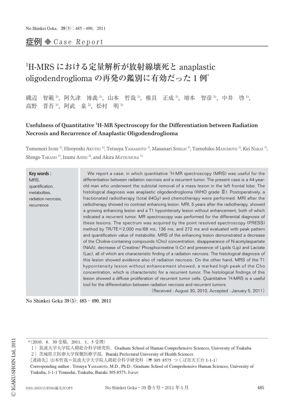
We report a case,in which quantitative 1H-MR spectroscopy (MRS) was useful for the differentiation between radiation necrosis and a recurrent tumor. The present case is a 44-year-old man who underwent the subtotal removal of a mass lesion in the left frontal lobe. The histological diagnosis was anaplastic oligodendroglioma (WHO grade Ⅲ). Postoperatively,a fractionated radiotherapy (total 64Gy) and chemotherapy were performed. MRI after the radiotherapy showed no contrast enhancing lesion. MRI,5 years after the radiotherapy,showed a growing enhancing lesion and a T1 hypointensity lesion without enhancement,both of which indicated a recurrent tumor. MR spectroscopy was performed for the differential diagnosis of these lesions. The spectrum was acquired by the point resolved spectroscopy (PRESS) method by TR/TE=2,000 ms/68 ms,136 ms,and 272 ms and evaluated with peak pattern and quantification value of metabolite. MRS of the enhancing lesion demonstrated a decrease of the Choline-containing compounds (Cho) concentration,disappearance of N-acetylaspartate (NAA),decrease of Creatine/ Phosphocreatine (t-Cr) and presence of Lipids (Lip) and Lactate (Lac),all of which are characteristic finding of a radiation necrosis. The histological diagnosis of this lesion showed evidence also of radiation necrosis. On the other hand,MRS of the T1 hypointensity lesion without enhancement showed,a marked high peak of the Cho concentration,which is characteristic for a recurrent tumor. The histological findings of this lesion showed a diffuse proliferation of recurrent tumor cells. Quantitative 1H-MRS is a useful tool for the differentiation between radiation necrosis and recurrent tumors.
Copyright © 2011, Igaku-Shoin Ltd. All rights reserved.