Japanese
English
- 有料閲覧
PPV購入案内
1,320円 (1,200円+税10%) こちらは、727ページから732ページの文献です。購入には医書.jp本会員登録が必要です。
会員登録完了後に改めて上記「購入サイトへ」を選択してください。
または「購入サイトへ」選択後に「購入ログイン」、「新規会員登録」の順に進んでください。
会員登録について詳しくはをご確認ください。
- Abstract 文献概要
- 1ページ目 Look Inside
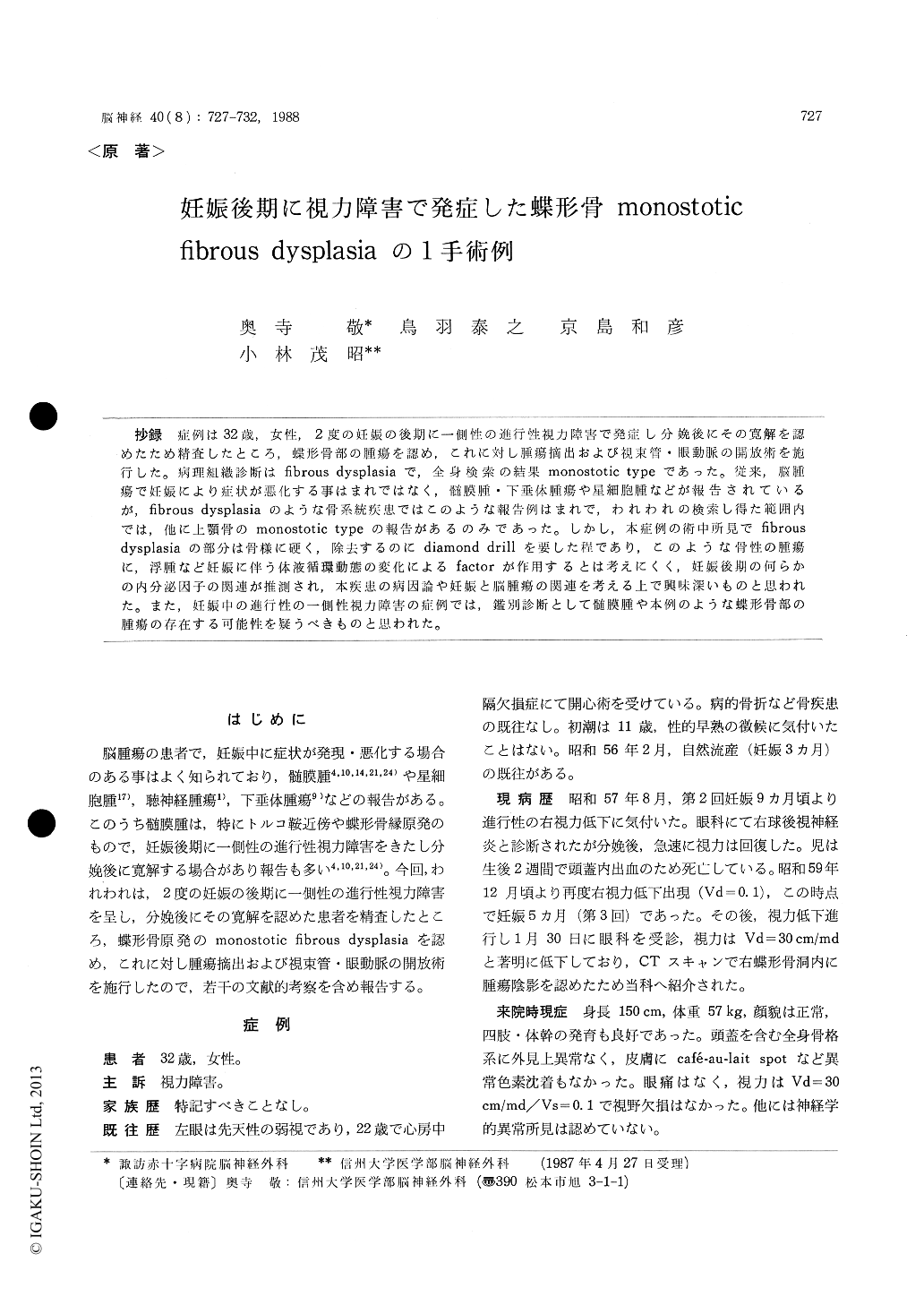
抄録 症例は32歳,女性,2度の妊娠の後期に一側性の進行性視力障害で発症し分娩後にその寛解を認めたため精査したところ,蝶形骨部の腫瘍を認め,これに対し腫瘍摘出および視束管・眼動脈の開放術を施行した。病理紅織診断はfibrous dysplasiaで,全身検索の結果monostotic typeであった。従来,脳腫瘍で妊娠により症状が悪化する事はまれではなく,髄膜腫・下垂体腫瘍や星細胞腫などが報告されているが,fibrous dysplasiaのような骨系統疾患ではこのような報告例はまれで,われわれの検索し得た範囲内では,他に上顎骨のmonostotic typeの報告があるのみであった。しかし,本症例の術中所見でfibrousdysplasiaの部分は骨様に硬く,除去するのにdiamond drillを要した程であり,このような骨性の腫瘍に,浮腫など妊娠に伴う体液循環動態の変化によるfactorが作用するとは考えにくく,妊娠後期の何らかの内分泌因子の関連が推測され,本疾患の病因論や妊娠と脳腫瘍の関連を考える上で興味深いものと思われた。また,妊娠中の進行性の一側性視力障害の症例では,鑑別診断として髄膜腫や本例のような蝶形骨部の腫瘍の存在する可能性を疑うべきものと思われた。
A case of monostotic fibrous dysplasia in the anterior skull base, which presented with visual disturbance during pregnancy, is reported.
A 32-year-old female was referred to our de-partment for examination of the progressing right visual disturbance in the third trimester of her second pregnancy. She had experienced the same episode in her first pregnancy, recovering from it after delivery. This time, however, the visual acuity did not change after delivery.
Plain craniograms showed sclerotic changes in the right sphenoidal ridge and the right frontal skull base. CT scan showed an isodense mass which was enhanced by contrast medium in the right sphenoidal sinus. Angiography demonstrated no positive findings. RI bone scintigram using 99 m Tc-MDP revealed an abnormal uptake in this region.
The patient was operated on in two stages. The first operation was transsphenoidal removal of the tumor in the sphenoidal sinus. The patho-logical diagnosis was fibrous dysplasia. Transfron-tal decompression of the right optic canal and ophthalmic artery was performed at the second operation. The tumor was totally removed and the decompressed orbital roof was reconstructed using an alumina ceramic plate. Visual acuity gradually improved in the follow-up study.
To our best knowledge, only one case of fibrous dysplasia with growth during pregnancy havebeen reported ; it was monostotic fibrous dyspla-sia of the maxilla. From the clinical course of our case, it is suggested that the pregnancy in-fluenced growth of the monostotic fibrous dyspla-sia ; possibly by way of hypothalamic hormonal factor.
Copyright © 1988, Igaku-Shoin Ltd. All rights reserved.