Japanese
English
- 有料閲覧
PPV購入案内
1,320円 (1,200円+税10%) こちらは、185ページから191ページの文献です。購入には医書.jp本会員登録が必要です。
会員登録完了後に改めて上記「購入サイトへ」を選択してください。
または「購入サイトへ」選択後に「購入ログイン」、「新規会員登録」の順に進んでください。
会員登録について詳しくはをご確認ください。
- Abstract 文献概要
- 1ページ目 Look Inside
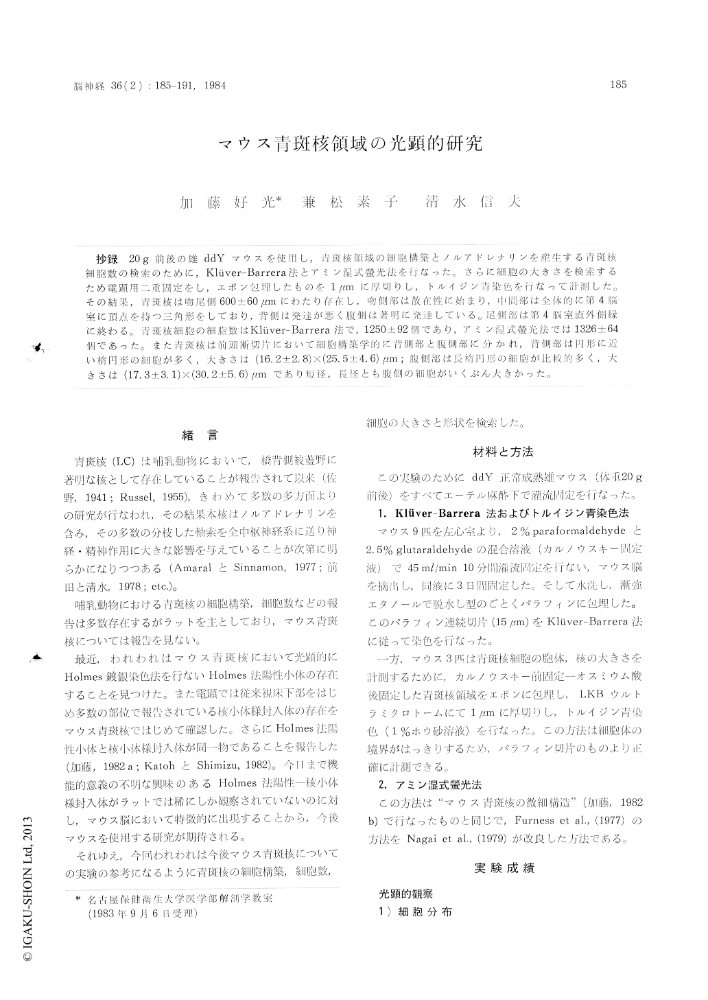
抄録 20g前後の雄ddYマウスを使用し,青斑核領域の細胞構築とノルアドレナリンを産生する青斑核細胞数の検索のために,Klüver-Barrera法とアミン湿式螢光法を行なった。さらに細胞の大きさを検索するため電顕用二重固定をし,エポン包埋したものを1μmに厚切りし,トルイジン青染色を行なって計測した。その結果,青斑核は吻尾側600±60μmにわたり存在し,吻側部は散在性に始まり,中間部は全体的に第4脳室に頂点を持つ三角形をしており,背側は発達が悪く腹側は著明に発達している。尾側部は第4脳室直外側縁に終わる。青斑核細胞の細胞数はKlüver-Barrera法で,1250±92個であり,アミン湿式螢光法では1326±64個であった。また青斑核は前頭断切片において細胞構築学的に青側部と腹側部に分かれ,背側部は円形に近い楕円形の細胞が多く,大きさは(16.2±2.8)×(25.5±4.6)μm;腹側部は長楕円形の細胞が比較的多く,大きさは(17.3±3.1)×(30.2±5.6)μmであり短径,長径とも腹側の細胞がいくぶん大きかった。
In an attempt to give some contribution to the morphology of locus coeruleus (LC) in the ddY mouse (weighing about 20 g), we made light-microscopic investigations using several staining methods in normal condition.
First, we studied the cytoarchitectonics and cell numbers of LC by the Kluver-Barrera method and amine demonstrating wet fluorescent method. LC of the mouse measured 600-1-60 pm in antero-posterior extent in the dorso-lateral tegmental area. At the rostral part of LC small number ofconstituent cells were disseminated, the middle part (principal part) showing the triangle-shaped cell accumulation with an apex directing to the IV ventricle. The caudal part forming small accumulation of cells ended directly beneath the IV ventricle.
Number of unilateral LC cells amounted to 1250 ±92 by Kluver-Barrera method, while 1326±44 by the wet fluorescent method. The LC was cytoarchitectonically divided into the dorsal and ventral parts. The cell size of the dorsal part averaged (16.2±2.8)×(25.5±4.6)μm along the longest and shortest axes, while that of ventral part (17.3±3.1)×(30.2±5.6)μm.
Copyright © 1984, Igaku-Shoin Ltd. All rights reserved.