Japanese
English
- 有料閲覧
- Abstract 文献概要
- 1ページ目 Look Inside
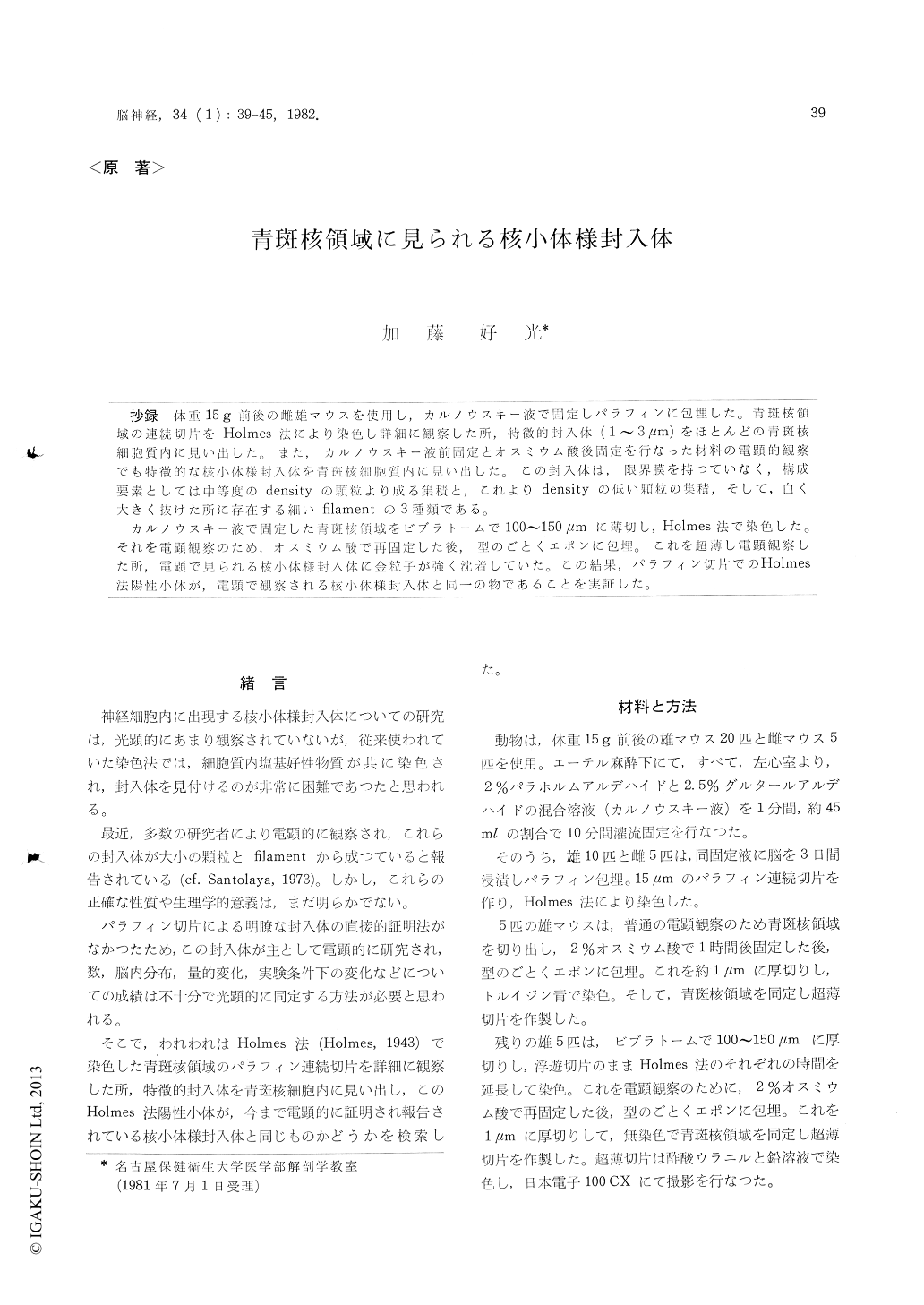
抄録 体重15g前後の雌雄マウスを使用し,カルノウスキー液で固定しパラフィンに包埋した。青斑核領域の連続切片をHolmes法により染色し詳細に観察した所,特徴的封入体(1〜3μm)をほとんどの青斑核細胞質内に見い出した。また,カルノウスキー液前固定とオスミウム酸後固定を行なった材料の電顕的観察でも特徴的な核小体様封入体を青斑核細胞質内に見い出した。この封入体は,限界膜を持つていなく,構成要素としては中等度のdensityの顆粒より成る集積と,これよりdensityの低い顆粒の集積,そして,白く大きく抜けた所に存在する細いfilamentの3種類である。
カルノウスキー液で固定した青斑核領域をビブラトームで100〜150μmに薄切し,Holmes法で染色した。それを電顕観察のため,オスミウム酸で再固定した後,型のごとくエポンに包埋。これを超薄し電顕観察した所,電顕で見られる核小体様封入体に金粒子が強く沈着していた。この結果,パラフィン切片でのHolmes法陽性小体が,電顕で観察される核小体様封入体と同一の物であることを実証した。
In the locus coeruleus of the normal mouse the presence and fine structure of nucleolus-like bodies have been studied by light and electron micro-scopic observation. In light microscopic observa-tion, it was clearly found that the bodies were characteristically stained for the first time in the mouse locus coeruleus by Holmes' silver staining. In the electron microscopic observation of conven-tional aldehyde-osmium fixed materials the locus coeruleus cell cytoplasm frequently showed the round nucleolus-like inclusion bodies of various size (1-3μm). These bodies devoid of the limiting membrane showed various size and structure: some bodies consist of complete aggregate of granules of medium-density, other bodies containing clear spaces of varying shape and size, which included accumulated fine filaments and round less dense granular body.
The present investigation is to determine whether or not the Holmes positive bodies are identical with electron microscopically demonstrable nucleo-lus-like bodies. Upon electron microscopic examina-tion of vibratome materials (100-150μm) treated by Holmes' method, we fortunately found the picture of nucleolus-like bodies loaded with heavy deposition of gold particles. From the observations stated above, we confirmed the identity of cyto-plasmic inclusion bodies stained by Holmes' method of paraffin sections with the nucleolus-like bodies demonstrated under electron microscope.
Therefore, though this silver metbod is devoid of histochemical values, it is worthwhile to clearly demonstrate the intracytoplasmic nucleolus-like bodies and their distribution and amount in the various parts of the brain. By the Holmes staining of the paraffin sections and electron microscopic observation, it was clarified that nucleolus-like bodies were present in almost all the locus coeruleus cell cytoplasm of both male and female mice.

Copyright © 1982, Igaku-Shoin Ltd. All rights reserved.


