Japanese
English
- 有料閲覧
PPV購入案内
1,320円 (1,200円+税10%) こちらは、205ページから214ページの文献です。購入には医書.jp本会員登録が必要です。
会員登録完了後に改めて上記「購入サイトへ」を選択してください。
または「購入サイトへ」選択後に「購入ログイン」、「新規会員登録」の順に進んでください。
会員登録について詳しくはをご確認ください。
- Abstract 文献概要
- 1ページ目 Look Inside
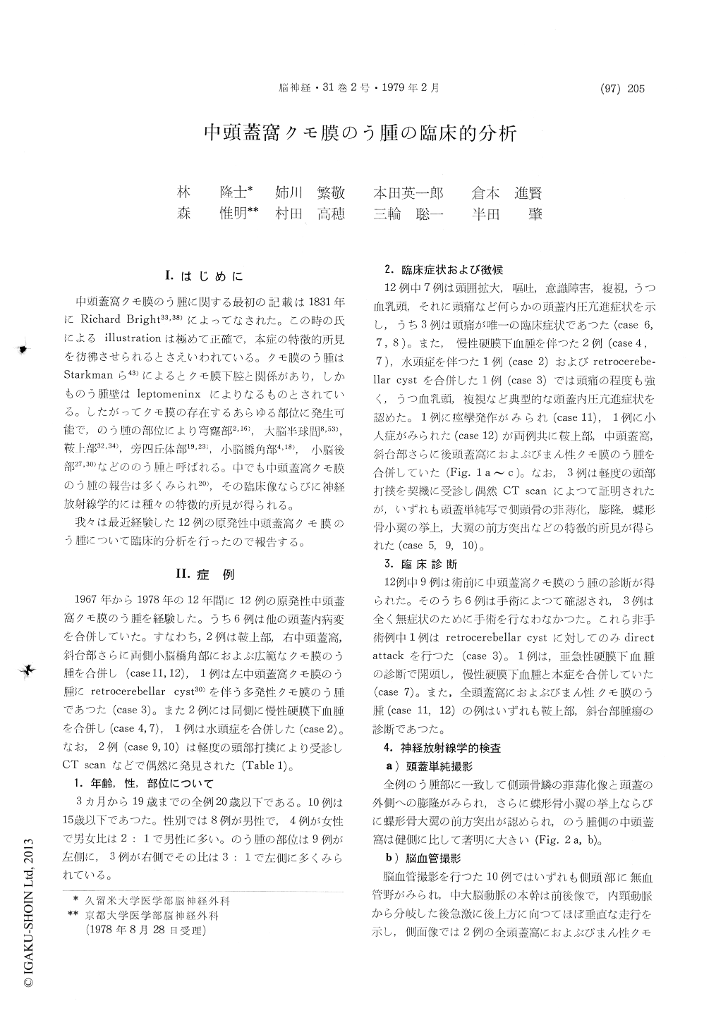
I.はじめに
中頭蓋窩クモ膜のう腫に関する最初の記載は1831年にRichard Bright33,38)によってなされた。この時の氏によるillustrationは極めて正確で,本症の特徴約所見を彷彿させられるとさえいわれている。クモ膜のう腫はStarkmanら43)によるとクモ膜下腔と関係があり,しかものう腫壁はleptomeninxによりなるものとされている。したがってクモ膜の存在するあらゆる部位に発生可能で,のう腫の部位により穹窿部2,16),大脳半球間8,53),鞍上部32,34),旁四丘体部19,23),小脳橋角部4,18),小脳後部27,30)などののう腫と呼ばれる。中でも中頭蓋窩クモ膜のう腫の報告は多くみられ20),その臨床像ならびに神経放射線学的には種々の特徴的所見が得られる。
我々は最近経験した12例の原発性中頭蓋窩クモ膜のう腫について臨床的分析を行ったので報告する。
Intraarachnoid cyst may occur in every site where the arachnoid exits, and it can be seen in the middle fossa, convexity, interhemisphere, parasellar, cerebellopontine angle and retrocerebellar regions. Among them, there is a high incidence of arachnoid cyst in the middle fossa, and is diversely charac-terized by its clinical symptoms and radiological findings. Further, arachnoid cyst in the middle fossa is associated with some agenetic state of the temporal lobe and its adjacent tissue. There are two theories on the pathogenesis of this cyst. One theory is that a localized defect of the temporal lobe is producted due to congenital anomary in opercularization, and an abnormal dilatation of the arachnoid space coinciding with the defective partoccurs, which forms a cyst after its loculation (subarachnoid cyst). The other theory is that an intra-arachnoid cyst is formed due to anomaly in systemic genetic process of the subarachnoid space, and because of the diturbance of opercularization, a localized arrest of the development of the temporal lobe is caused.
This paper describes the clinical analyses and radiological findings of 12 cases of " idiopathic" arachnoid cysts in the middle fossa encountered during the past 12 years. Their results were following.
1. The ages of the patients in 10 cases out of 12 cases were under 15 years of age, and 8 cases were males. The site of the cyst was on the left side in 9 cases.
2. On the plain skull radiography, thinning and bulging of the temporal bone at the lesion, elevation of the lesser wing of the sphenoid, and forward projection of the greater wing of the sphenoid were observed in all cases.
3. On cerebral angiography, a characteristic elevation of the middle cerebral artery and hypo-plasty of the insular portion and opercular portion were observed; and on venous phase, a defect of the middle cerebral vein, backward deviation of the Labbe' vein, and elevation of the Rosenthal vein were seen.
4. On Amipaque CT cisternography, there were two types, one was communicable and other was non-communicable between the cyst and its adjacent subarachnoid space.
Of the 12 cases the authors experienced there were those with diffuse arachnoid cyst extending to the whole skull base and those with retro-cerebellar cyst. Also the authors observed other intracranial disease such as chronic subdural hematoma and hydrocephalus in the arachnoid cysts.
Copyright © 1979, Igaku-Shoin Ltd. All rights reserved.