Japanese
English
- 有料閲覧
- Abstract 文献概要
- 1ページ目 Look Inside
I.はじめに
今回は第1報の結果にもとづき,各時期の実験水頭症犬における硬膜外圧測定を行ない,脳室拡大ならびに,CT上のperiventricular lucency (PVL)15)と頭蓋内圧との関係について検討した。
Epidural pressure (EDP) were monitored by Ladd intracranial pressure monitoring apparatus in the various stages of kaolin-induced canine hydro-cephalus, and computed tomopraghy (CT) were perf-ormed at the same time for observations of the ve-ntricles. On the measurement of EDP, the experi-mental dogs were anesthesized with intravenous injection of Pentobarbital Sodium, and were immobi-lized and put on controlled respiration with musclerelaxant. During the measurements, room temperature and body temperature of dogs were kept at the constant ranges, and blood pressure, rates and tidal volume of respirations and arterial blood gas analysis were checked frequently. EDP was monitored for at least 1 hour, and in some dogs, for 24 hours especially immediately after kaolin injection. The average of EDP in normal 8 dogs was 9.1 ±2.0 cmH2O. In 24 hours after kaolin injection EDP increased in all dogs, but is showed remarkable individual variations.
CT scans were performed in 1 day to 20 weeks after kaolin injection with the same method as in part 1. In addition to the previous observations, the 4th ventricle was examined in the present study.
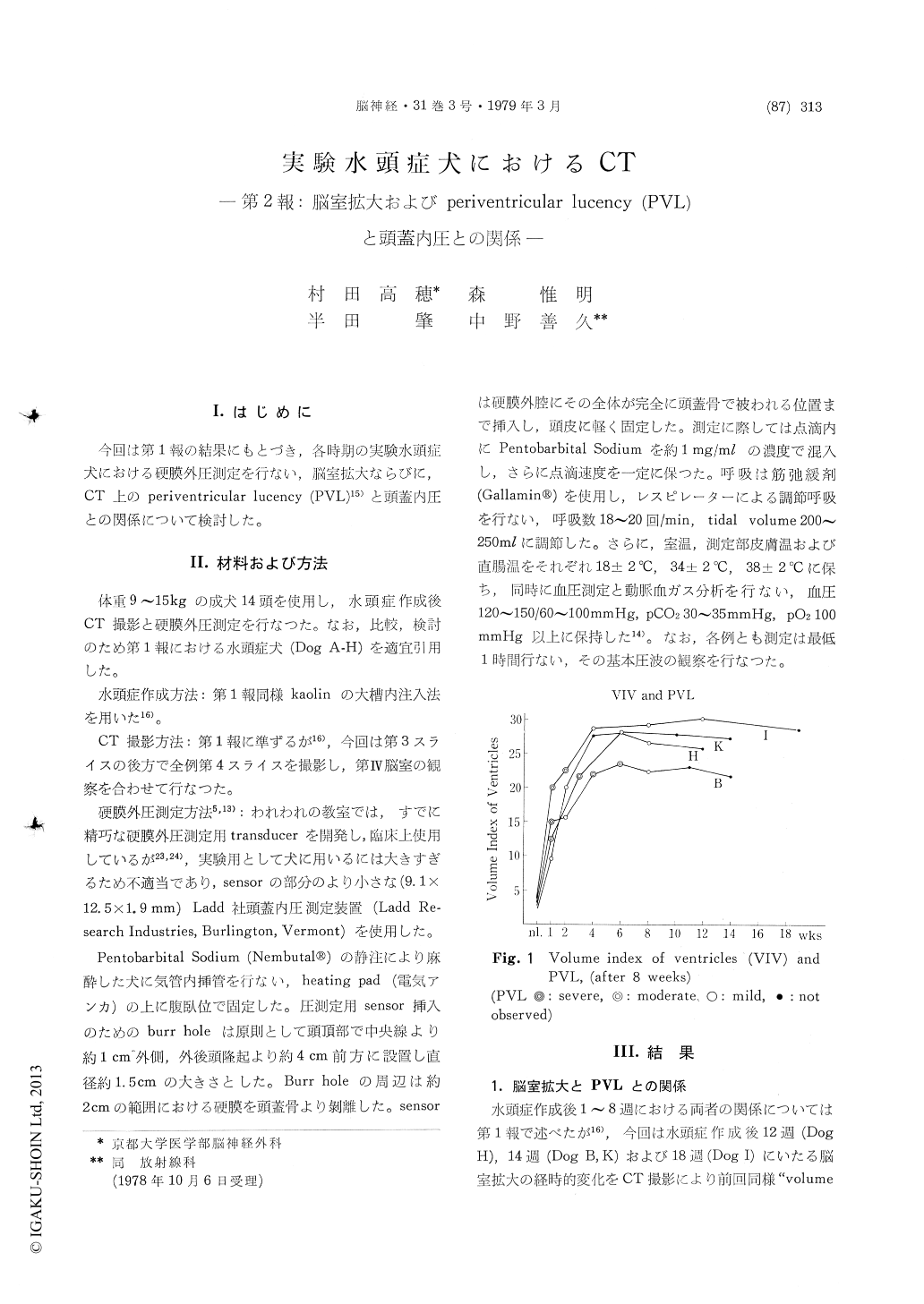
According to these results, in 5 of 8 dogs, intra-ventricular pressure (IVP) that was equivalent to EDP, became maximum in 1 week (24.3 5.7 cmH2O in EDP) and then decreased quickly in 2 to 8 weeks. In 8 weeks IVP returned to normal or even to subnormal ranges (7.8 ±1.2 cmH20 in EDP). There was a correlation between the in-creased IVP that preceded the ventricular en-largement, and the degree of the ventricularenlargement. In an acute stage, periventricular lucency (PVL) had a tendency to appear when IVP increased (more than 19. 5 cmH2O in EDP), and there was a correlation between the degree of PVL and the increase of IVP. Consequently, PVL in acute stage was considered to be a pressure sign on CT scans. However, PVL in a chronic or non-hypertensive stages might not be caused by a periventricular edema due to increased IVP. We considered it to be infarction or necrosis of the periventricular white matter that is a late effect of circulatory disturbances in a hypertensive stage. Pathogenesis of PVL in a chronic stage will be discussed in the next paper (part 3).
In 3 of 8 dogs IVP did not increase in 1 to 2 weeks, and it reached maximum in 4 to 6 weeks along with ventricular enlargement and mild or moderate PVL on CT. In kaolin-induced hydro-cephalus, various types of hydrocephalus were encountered ; obstructive, communicating and mixed types. It is conceivable that these different types of hydrocephalus have been induced by the differ-ence of the site and degree of obstruction caused by kaolin injection.

Copyright © 1979, Igaku-Shoin Ltd. All rights reserved.


