Japanese
English
- 有料閲覧
- Abstract 文献概要
- 1ページ目 Look Inside
I.はじめに
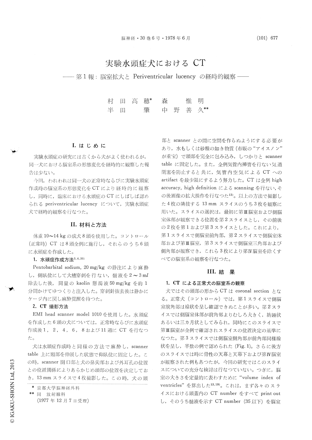
実験水頭症の研究には古くから犬がよく使われるが,同一犬における脳室系の形態変化を経時的に観察した報告は少ない。
今回,われわれは同一犬の正常時ならびに実験水頭症作成時の脳室系の形態変化をCTにより経時的に観察し,同時に,臨床における水頭症のCTにしばしば認められるperiventricular lucencyについて,実験水頭症犬で経時的観察を行なつた。
There have been few reports of observation on changes of ventricular size and shape on identical dogs during development of experimental hydro-cephalus. We studied the morphological changes of ventricles in experimental hydrocephalus on identical dogs by successive computed tomography(CT).
CT scans on dogs were performed in the coronal sections because of the shape of the head. In the normal ventricles, the frontal and temporal horns were slit-like and not always possible to recognize, and the body was spindle or crescent shaped and easily identified. In the same slice where the body was identified, the third ventricle was recognized in all cases.
Hydrocephalus was induced by cisternal injection of kaolin suspension. CT examinations were done on identical dogs before and on 1,2,4,6,8 and 11 weeks after kaolin injection.
In these dogs, the degrees of ventricular en-largement were studied by "volume index of ven-tricles" which was calculated from printed out CT numbers. The degree of ventricular enlargement was most remarkable in the first week and less remarkable in the second week. The ventricles reached the maximum size in the fourth to sixthweek and seemed to reduce slightly in size after the eighth week.
Periventricular lucency on CT was also detected in the experimental canine hydrocephalus. It was always most remarkable in the frontal horn of lateral ventricles, and differed in degree from severe to mild. The severe case showed irregular land horn-shaped lucency in the periventricular regions. The periventricular lucency was distinct in the first week after kaolin injection. It gradually became indistinct and had a tendency to disappear thereafter. We consider that the periventricular lucency in the acute stage may represent peri-ventricular edema causod by an increase in water content in the periventricular white matter.
CT examinations on more chronic hydrocephalus and after a shunting operation, and a correlation between intrancranial pressure and periventricular lucency are now under investigation.

Copyright © 1978, Igaku-Shoin Ltd. All rights reserved.


