Japanese
English
- 有料閲覧
PPV購入案内
1,320円 (1,200円+税10%) こちらは、1209ページから1214ページの文献です。購入には医書.jp本会員登録が必要です。
会員登録完了後に改めて上記「購入サイトへ」を選択してください。
または「購入サイトへ」選択後に「購入ログイン」、「新規会員登録」の順に進んでください。
会員登録について詳しくはをご確認ください。
- Abstract 文献概要
- 1ページ目 Look Inside
I.はじめに
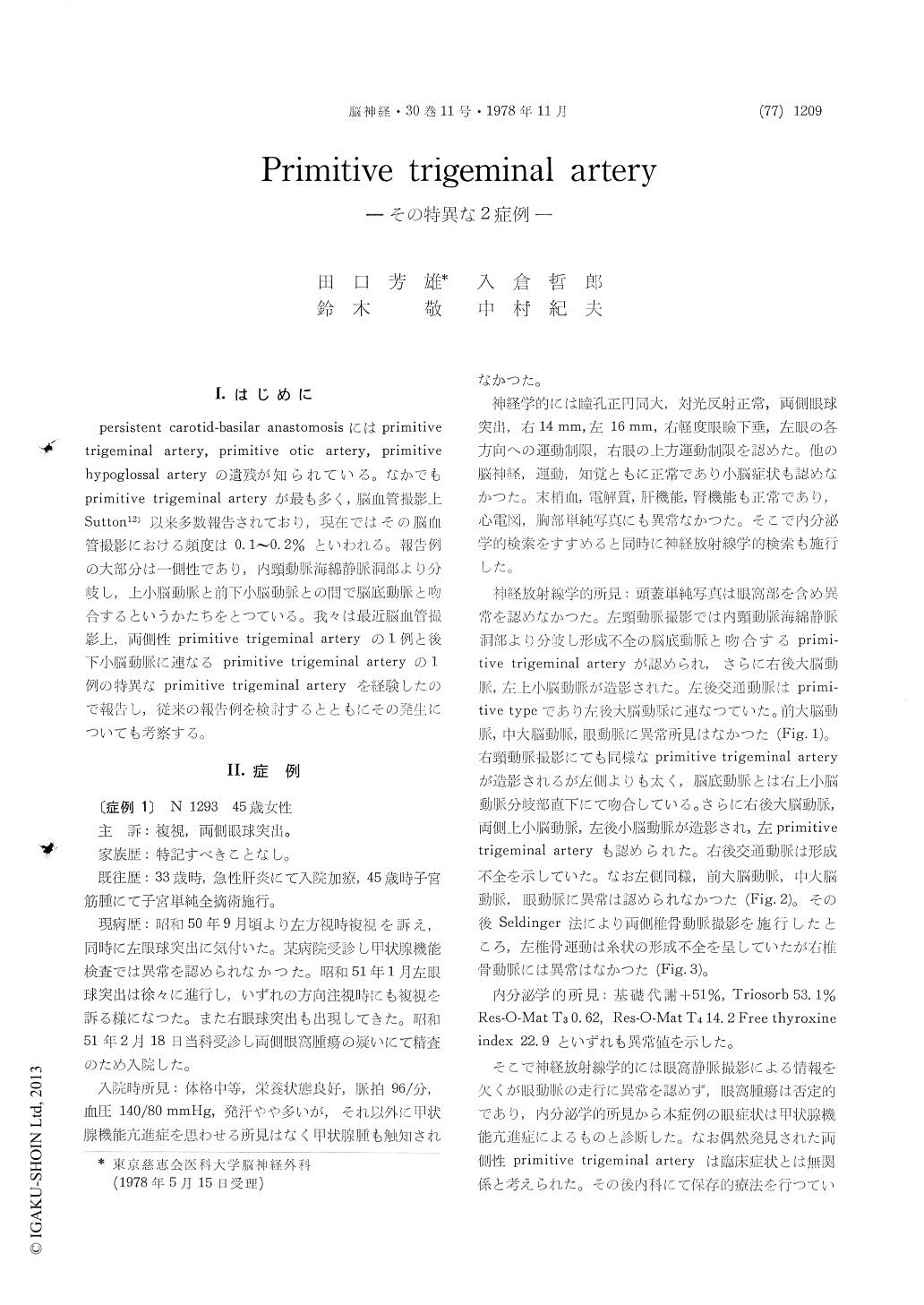
persistent carotid-basilar anastomosisにはprimitivetrigeminal artery,primitive otic artery,primitivehypoglossal arteryの遺残が知られている。なかでもprimitive trigeminal arteryが最も多く,脳血管撮影上Sutton12)以来多数報告されており,現在ではその脳血管撮影における頻度は0.1〜0.2%といわれる。報告例の大部分は一側性であり,内頸動脈海綿静脈洞部より分岐し,上小脳動脈と前下小脳動脈との間で脳底動脈と吻合するというかたちをとつている。我々は最近脳血管撮影上,両側性primitive trigeminal arteryの1例と後下小脳動脈に連なるprimitive trigeminal arteryの1例の特異なprimitive trigeminal arteryを経験したので報告し,従来の報告例を検討するとともにその発生についても考察する。
The primitive trigeminal artery is the most common of the three well known persistent carotid-basilar anastomoses. Recently two cases of unusual primitive trigeminal artery were encountered in the neurosurgical department of Jikei UniversityHospital. The first was bilateral primitive trigeminal arteries accompanied by hyperthyroidism. The second was a primitive trigeminal artery connecting to the ipsilateral posterior inferior cerebellar artery as a loop in association with a tuberculum sellae meningioma.
Case 1. A 45 year-old women was admitted for evaluation of bilateral exophthalmos of five month duration. Physical examination revealed no struma. Positive findings on neurological examination included bilateral exophthalmos and limitation of ocular movement to any direction. Pupillary signs were normal. Carotid angiogram revealed no ab-normalities except for bilateral primitive trigeminal arteries connecting between the cavernous portion of the internal carotid arteries and the basilar artery.
Endocrinological examination suggested hyper-thyroidism. So she was diagnosed as bilateral exophthalmos due to hyperthyroidism. Bilateral primitive trigeminal arteries were coincidental.
Case 2. A 46 year-old man was first seen with complaint of decreased visual acuity of one year duration. Positive findings on neurological testing included impairment of visual acuity and bitemporal hemianopia. Motor and sensory examinations were normal. Plain craniogram disclosed typical blistering of the sphenoid sinus. Carotid angiogram showed large tumor stain in the suprachiasmatic region. Furthermore a right carotid angiogram revealed a right primitive trigeminal artery running into the posterior fossa without connection to the basilar artery and supplying a tributary of the ipsilateral posterior inferior cerebellar artery mainly. He was diagnosed as tuberculum sellae meningioma and the primitive trigeminal artery was coincidental. A bifrontal craniotomy was performed with total removal of the elastic hard mass. Microscopic analysis disclosed meningotherial meningioma with psammoma body.
The angiographic incidence of the primitive trigeminal artery is approximately O.1% to 0.2% and 0.8% in our hospital. However, there are only two cases of bilateral primitive trigeminal arteries in the literature and only few cases were reported which the internal carotid artery supplys the posterio inferior cerebellar artery distribution directly through a primitive trigeminal artery. From on embryological point of view, these two cases contribute various interesting suggestions to the development of cerebrovascular distributions.
Copyright © 1978, Igaku-Shoin Ltd. All rights reserved.