Japanese
English
- 有料閲覧
PPV購入案内
1,320円 (1,200円+税10%) こちらは、309ページから315ページの文献です。購入には医書.jp本会員登録が必要です。
会員登録完了後に改めて上記「購入サイトへ」を選択してください。
または「購入サイトへ」選択後に「購入ログイン」、「新規会員登録」の順に進んでください。
会員登録について詳しくはをご確認ください。
- Abstract 文献概要
- 1ページ目 Look Inside
I.はじめに
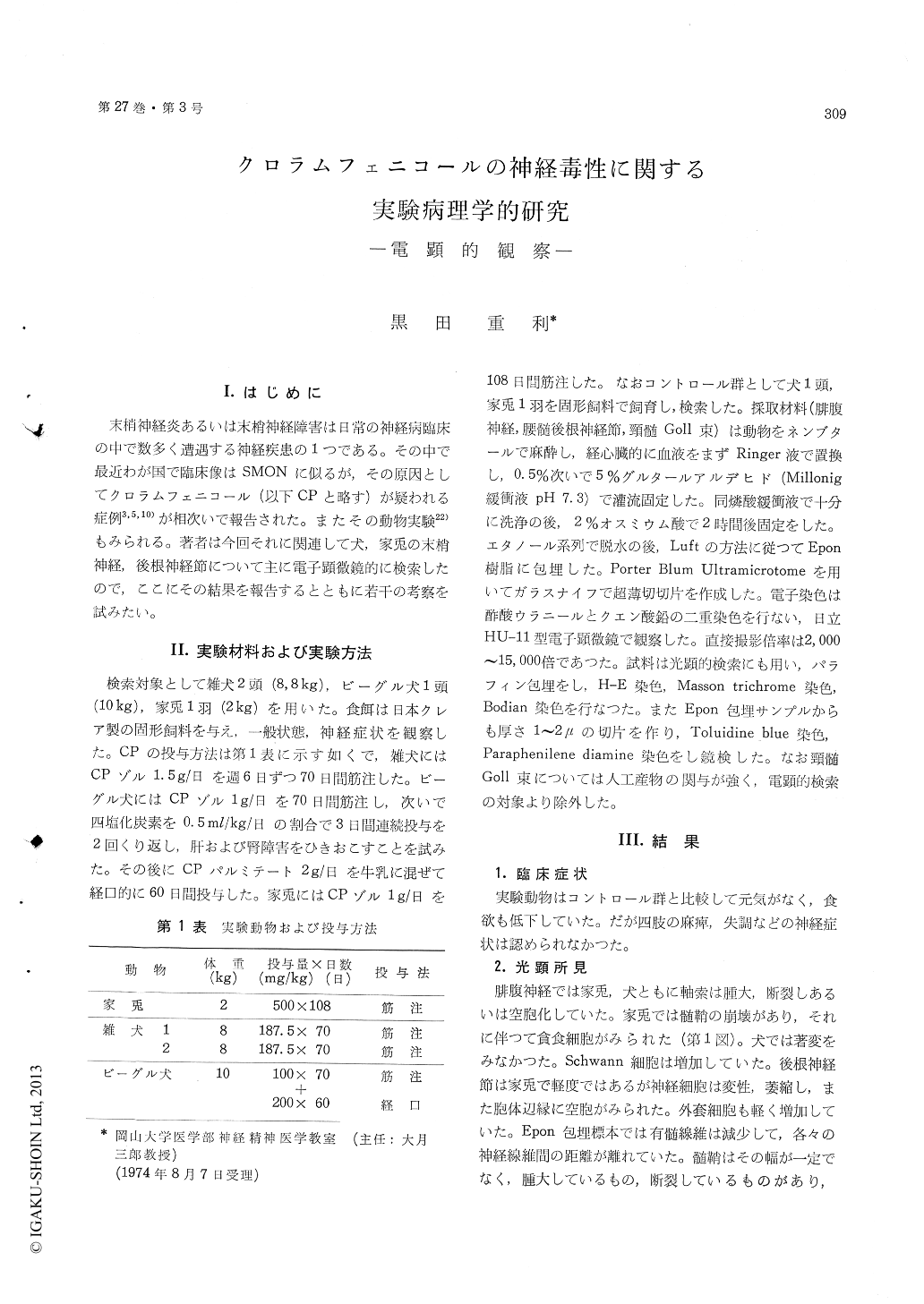
末梢神経炎あるいは末梢神経障害は日常の神経病臨床の中で数多く遭遇する神経疾患の1つである。その中で最近わが国で臨床像はSMONに似るが,その原因としてクロラムフェニコール(以下CPと略す)が疑われる症例3,5,10)が相次いで報告された。またその動物実験22)もみられる。著者は今回それに関連して犬,家兎の末梢神経,後根神経節について主に電子顕微鏡的に検索したので,ここにその結果を報告するとともに若干の考察を試みたい。
We performed animal experiments on neuropathy that was recently considered to be induced by chloramphenicol (CP) in Japan, mainly by electron microscopy. The results are briefly summarized as follows.
1) Into two mongrel dogs 1.5 g/day of CP was injected for 70 consecutive days, and into one rabbit1.0 g/day of it for 108 days intramuscularly. A beagle was at first given 1.0 g/day of CP for 70 days intramuscularly, followed by carbon tetra-chloride administration, then 2.0 g/day of CP pal-mitate was administered orally for 60 days. As a result no distinct neuropathy was observed. Each of these animals was then given perfusion fixation, and sural nerves and lumbar posterior ganglion cells from these animals were studied by light microscopy and electron microscopy. One dog and one rabbit each served for control.
2) With sural nerve changes of myelin sheaths and Schwann cells were prominent, revealing the swelling and enlargement of the myelin sheath or disintegration, thinning and honeycomb-like struc-ture, Myelin ovoid structures could also be observed. Schwann cells were enlarged and increased in number, and as for the intracellular changes there was observed an increase of endoplasmic reticuli, dense bodies and vacuoles. There could also be seen onion-bulb formation. The nerve axon showed an increase of neurofilaments and accumulation of mitochondria and there could be observed the pro-jection of the axon-Schwann membrane into nerve axon.
3) Nerve cells in lumbar root ganglia showed various changes. The cisternae of endoplasmic reticuli were irregularly swollen, their ribosomes no longer were in the rosette formation, and their number was decreased and some of them showed small granules scattered in the cytoplasm as spots. Mitochondria have undergone a marked change ; many of them showing the direction of cristae de-ranged and became lost, or some swollen into a vacuolar formation. Sometimes neurofilaments were decreased in number, or some of them were in-creased. The nucleus showed indentations of the nuclear membrane, and in such an instance, nuclear pores were seen densely pached on the membrane surface, and some lumina of the nuclear membrane were dense. The lining cells showed no positive pathological changes.
Copyright © 1975, Igaku-Shoin Ltd. All rights reserved.