Japanese
English
- 有料閲覧
PPV購入案内
1,320円 (1,200円+税10%) こちらは、831ページから839ページの文献です。購入には医書.jp本会員登録が必要です。
会員登録完了後に改めて上記「購入サイトへ」を選択してください。
または「購入サイトへ」選択後に「購入ログイン」、「新規会員登録」の順に進んでください。
会員登録について詳しくはをご確認ください。
- Abstract 文献概要
- 1ページ目 Look Inside
I.はじめに
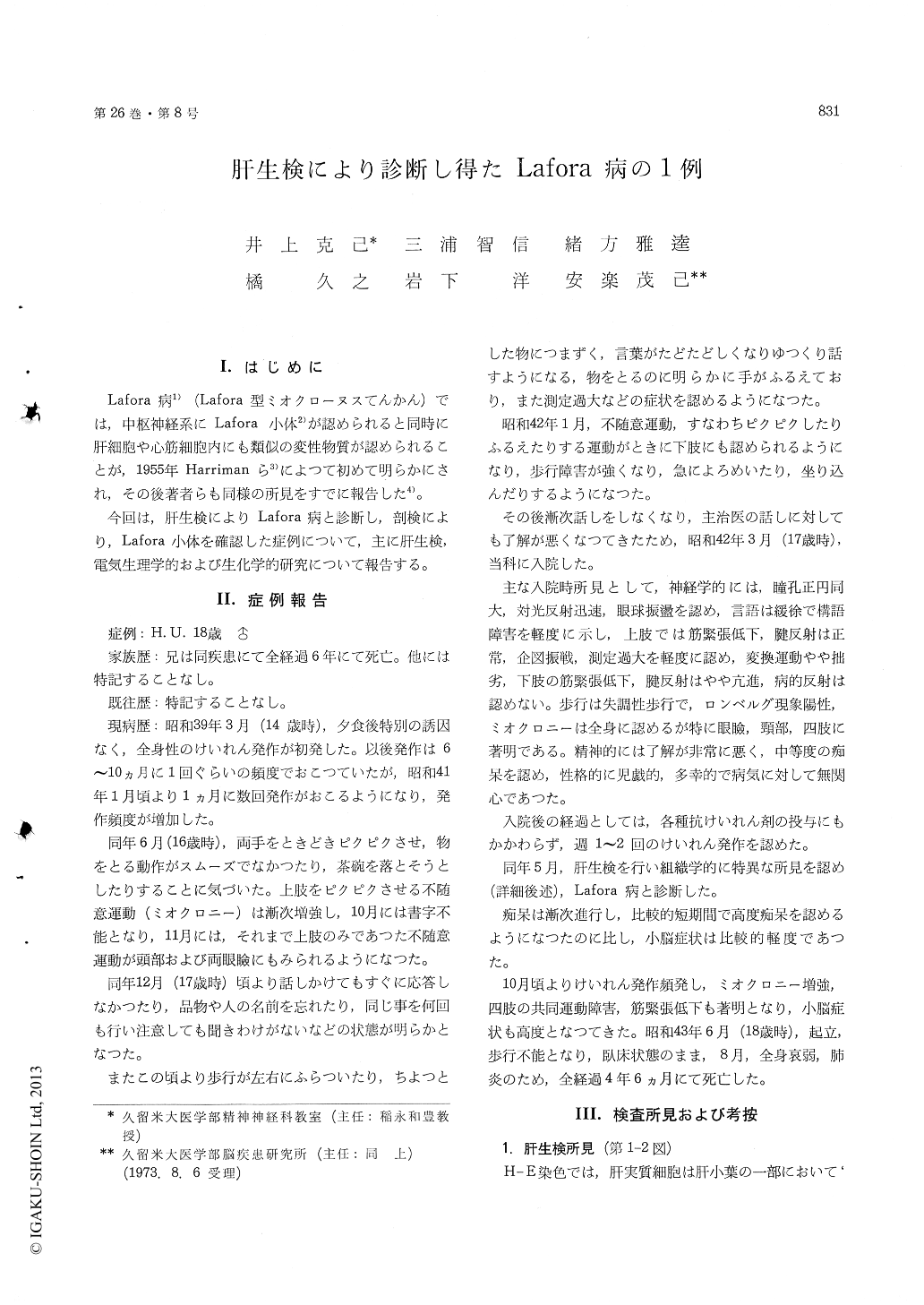
Lafora病1)(Lafora型ミオクローヌスてんかん)では,中枢神経系にLafora小体2)が認められると同時に肝細胞や心筋細胞内にも類似の変性物質が認められることが,1955年Harrimanら3)によって初めて明らかにされ,その後著者らも同様の所見をすでに報告した4)。
今回は,肝生検によりLafora病と診断し,剖検により,Lafora小体を確認した症例について,主に肝生検,電気生理学的および生化学的研究について報告する。
The case, an 18-year-old man whose elder brother also had died of the same disease, was seized with a cramp at 14. Two years later myoclonus appeared and the patient was admitted to our hospital under the conditions of dementia and cerebellar symptoms at 17. Under the treatment for Lafora's disease diagnosed from the findings of liver biopsy, he died of marasmus 4 and a half years after onset of his illness. Postmortem examination revealed the pre-sence of Lafora bodies over the whole brain, which justified the diagnosis of Lafora's disease made athis living time.
Histological findings of liver biopsy as well as electrophysiological and biochemical findings of the present case are as follows :
1) Histological findings of liver biopsy : a part or the whole of the cytoplasm of liver cells was stained homogeneously and light-basically by H. E. staining and PAS-positive sediments were seem in accord with these sites.
2) Electrophysiological findings :
i) As to the relationship of changes in clinical symptoms with the EEG, in the period of convul-sion or myoclonus EEG showed polys-pikes, a poly-spike and wave complex, and paroxysmal high voltage slow wave bursts. These abnormal patterns were markedly activated by photic stimulation. After the appearance of dementia and cerebellar symptoms background activity changed to slower activity with lower voltage and became dysrhyth-mic. At terminal stage slowing of the background activity advanced increasingly, poly-spikes and a poly-spike and wave complex decreased, and then voltage of spike components became lower.
ii) All-night EEG recording of this case showed a prolonged sleep latency, an increasing awaking stage with no light sleep stage, and a decreasing paradoxycal sleep stage as compared with normal subjects. On the other hand, in contrast with the degenerative type of myolonus-epilepsy a remark-able difference was noted between the light and deep sleep stages in this case. The appearance of burst waves at the time of sleep decreased in fre-quency as the sleep became deeper. Above all it decreased conspicuously at the stage of paradoxycal sleep.
iii) Visually evoked response and photopalpebral reflex of the patient showed a markedly increased amplitude of the components compared with normal subjects or the degenerative type of myoclonus epilepsy.
3) Examination of acid mucopolysaccharides (AMPS) in the urine revealed :
i) In the group of Lafora's disease (2 cases), AMPS showed a tendency to increase. in compari-son with normal subjects or the degenerative type of myoclonus epilepsy.
ii) At electrophoresis, only in the group of Lafora's disease pattern corresponding to Hyaluronic acid were noted.
Copyright © 1974, Igaku-Shoin Ltd. All rights reserved.