Japanese
English
- 有料閲覧
PPV購入案内
1,320円 (1,200円+税10%) こちらは、1095ページから1099ページの文献です。購入には医書.jp本会員登録が必要です。
会員登録完了後に改めて上記「購入サイトへ」を選択してください。
または「購入サイトへ」選択後に「購入ログイン」、「新規会員登録」の順に進んでください。
会員登録について詳しくはをご確認ください。
- Abstract 文献概要
- 1ページ目 Look Inside
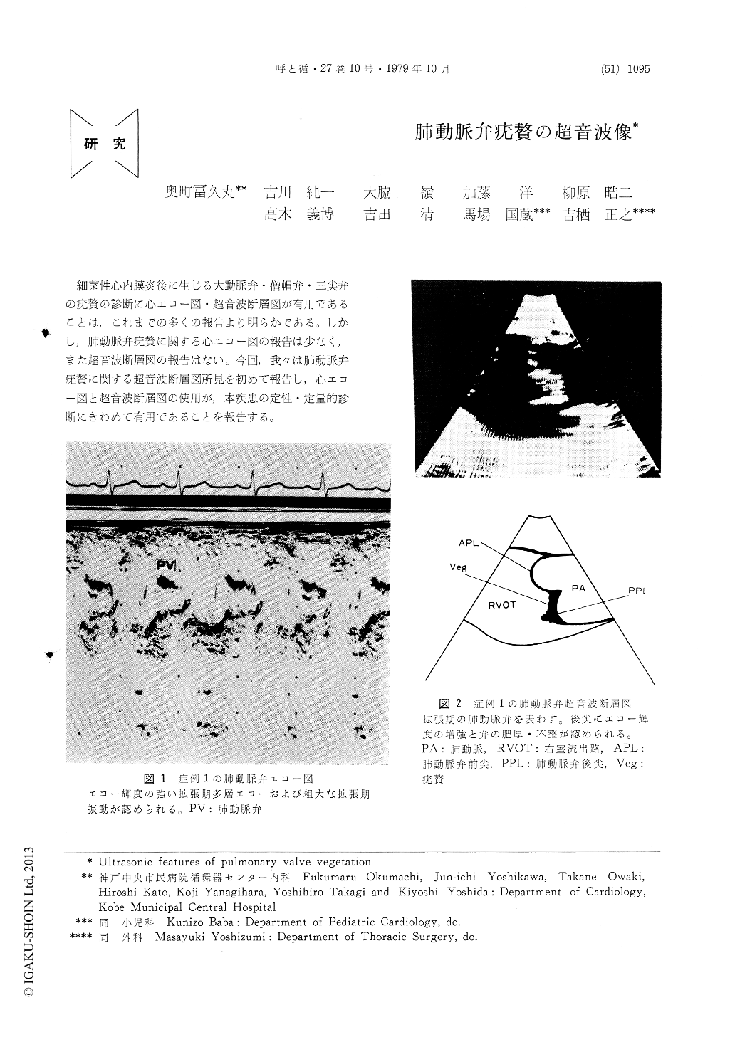
細菌性心内膜炎後に生じる大動脈弁・僧帽弁・三尖弁の疣贅の診断に心エコー図・超音波断層図が有用であることは,これまでの多くの報告より明らかである。しかし,肺動脈弁疣贅に関する心エコー図の報告は少なく,また超音波断層図の報告はない。今回,我々は肺動脈弁疣贅に関する超音波断層図所見を初めて報告し,心エコー図と超音波断層図の使用が,本疾患の定性・定量的診断にきわめて有用であることを報告する。
Real-time, cross-sectional echograms and M-mode echocardiograms of the pulmonary valve were obtained in 2 patients with pulmonary valve vegetation. The M-mode echocardiograms disclosed shaggy echoes in the region of the pulmonary valve in diastole in both cases. The real-time cross-sectional echograms demonstrated the size, location and motion of the vegetation in each case. Furthermore, in one case the flail pulmonary valve leaflets confirmed by operation was shown in the cross-sectional echograms. Cross-sectional echocardiography offers a direct, noninvasive method for visualizing pulmonary valve vegetation and resultant valve change, and should be an improvement over the indirect M-mode data.
Copyright © 1979, Igaku-Shoin Ltd. All rights reserved.