Japanese
English
- 有料閲覧
- Abstract 文献概要
- 1ページ目 Look Inside
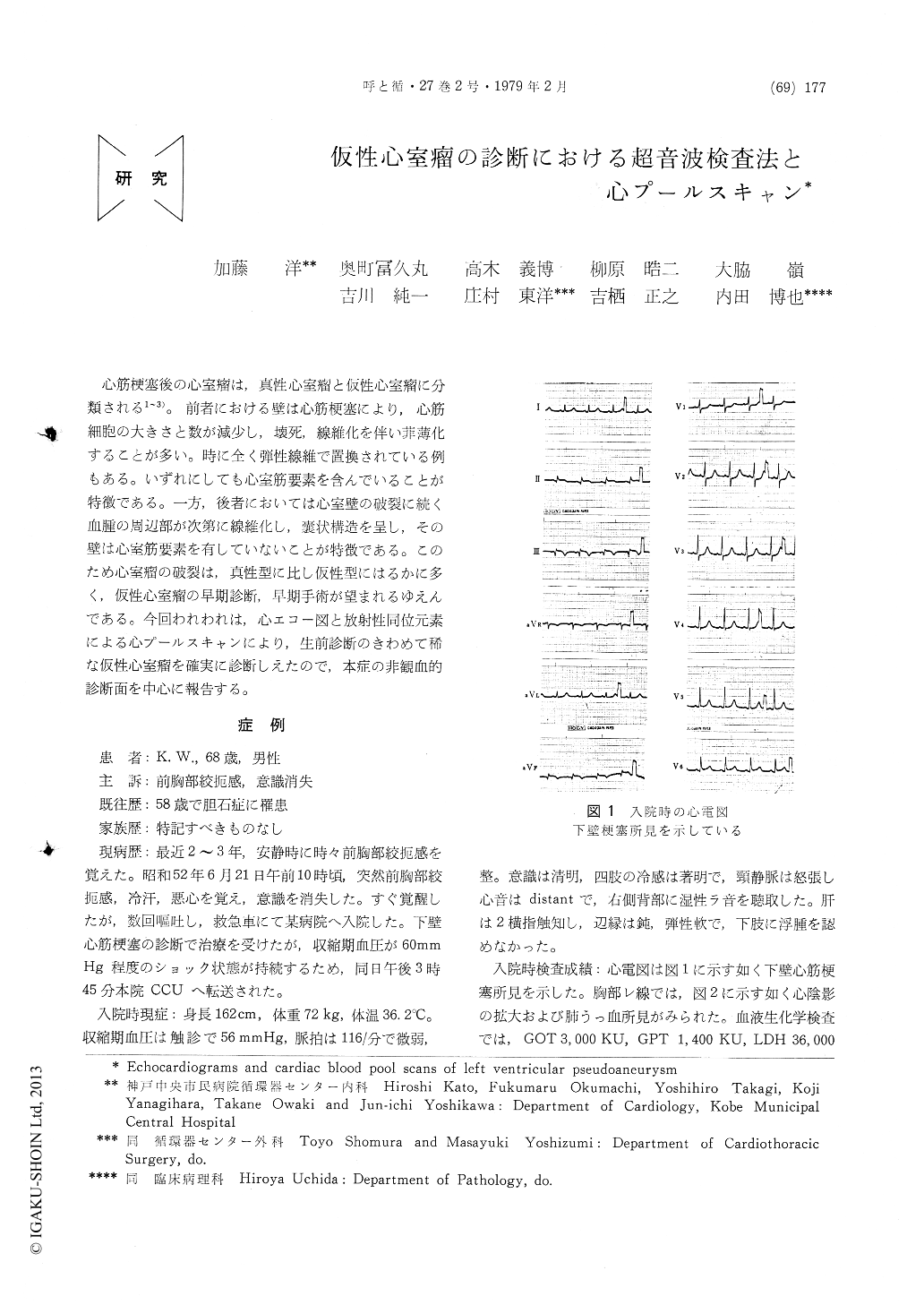
心筋梗塞後の心室瘤は,真性心室瘤と仮性心室瘤に分類される1〜3)。前者における壁は心筋梗塞により,心筋細胞の大きさと数が減少し,壊死,線維化を伴い菲薄化することが多い。時に全く弾性線維で置換されている例もある。いずれにしても心室筋要素を含んでいることが特徴である。一方,後者においては心室壁の破裂に続く血腫の周辺部が次第に線維化し,嚢状構造を呈し,その壁は心室筋要素を有していないことが特徴である。このため心室瘤の破裂は,真性型に比し仮性型にはるかに多く,仮性心室瘤の早期診断,早期手術が望まれるゆえんである。今回われわれは,心エコー図と放射性同位元素による心プールスキャンにより,生前診断のきわめて稀な仮性心室瘤を確実に診断しえたので,本症の非観血的診断面を中心に報告する。
The echocardiograms and cardiac blood poolscans were obtained from a patient with post-infarction pseudoaneurysm. The interruption of the posterior left ventricular wall, echo free space and the dense pericardium-like echo moving outward in systole were observed on the echocardiograms. The interruption of the posterior left ventricular wall was thought to reflect that the ultrasound beam passed through the opening between the left ventricle and pseudoaneurysm. The dense pericardium-like echo may be the peri-cardium and/or the wall of the pseudoaneurysm. Cardiac blood pool scanning demonstrated the saccular aneurysm which was separated by the left ventricle. Noninvasive methods including echocardiography and cardiac blood pool scanning seem to be safe and specific methods for the diagnosis of left ventricular pseudoaneurysm.

Copyright © 1979, Igaku-Shoin Ltd. All rights reserved.


