Japanese
English
- 有料閲覧
- Abstract 文献概要
- 1ページ目 Look Inside
左房粘液腫の心エコー図診断については,1959年Effertら1)が報告して以来,数多くの報告2〜5)がみられる。本症の特徴的所見として,拡張期に僧帽弁前尖の背方に出現する点状または苺状の異常エコーが知られている。一方,大動脈背方の左房における腫瘍エコーの出現は,腫瘍の可動性や大きさによって影響を受ける。腫瘍が小さく可動性が良好な場合,腫瘍エコーは全く認められず,腫瘍が増大し可動性が不良になるにしたがい,腫瘍エコーはまず収縮期のみに,ついで心周期を通じて左房内に出現するようになる6)。
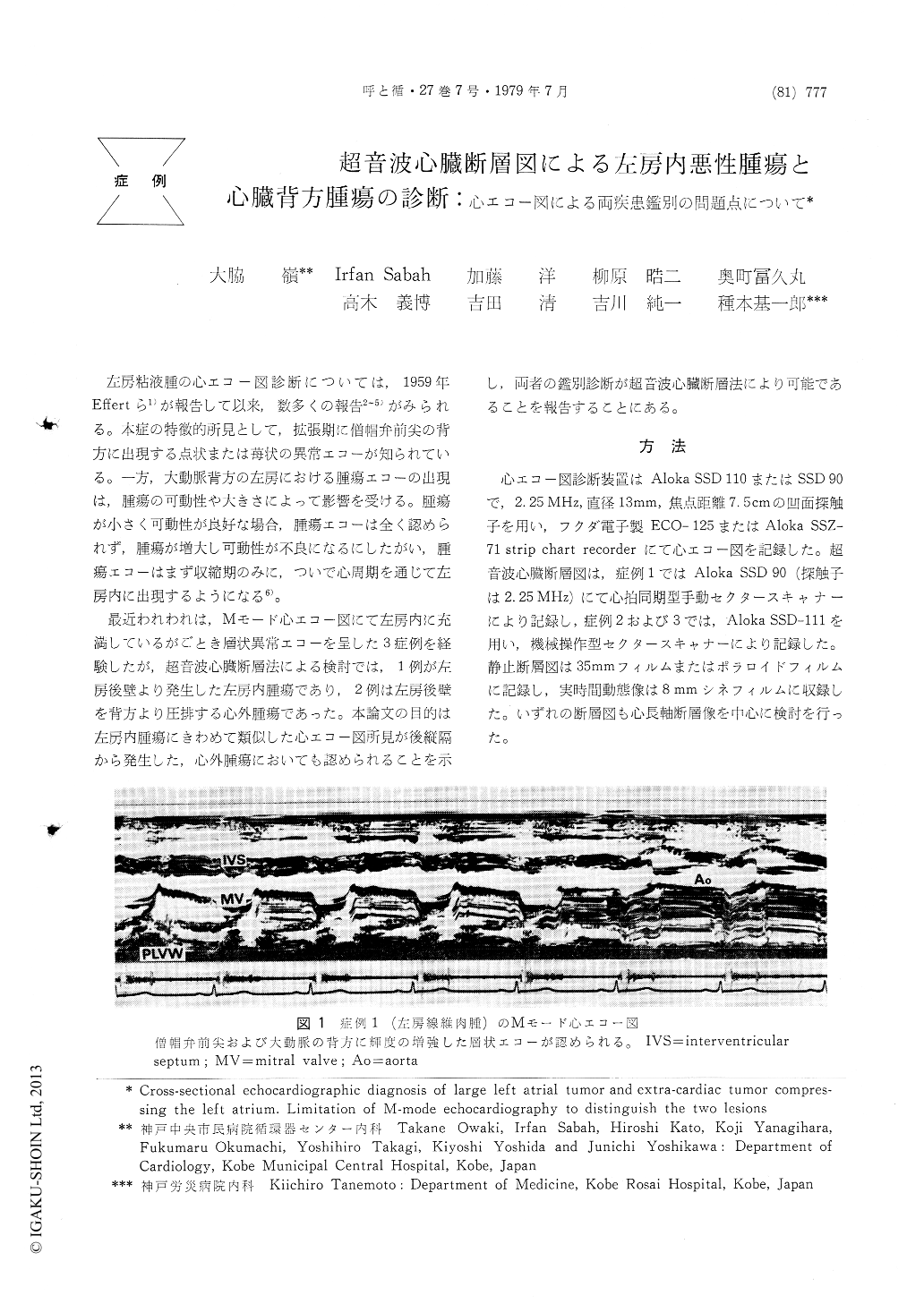
最近われわれは,Mモード心エコー図にて左房内に充満しているがごとき層状異常エコーを呈した3症例を経験したが,超音波心臓断層法による検討では,1例が左房後壁より発生した左房内腫瘍であり,2例は左房後壁を背方より圧排する心外腫瘍であった。本論文の目的は左房内腫瘍にきわめて類似した心エコー図所見が後縦隔から発生した,心外腫瘍においても認められることを示し,両者の鑑別診断が超音波心臓断層法により可能であることを報告することにある。
A case with left atrial fibrosarcoma arising from the posterior left atrial wall and two cases with extra-cardiac tumor compressing the left atrium were studied by M-mode echocardiography and cross-sectional echocardiography. In all cases, M-mode echocardiography revealed a mass of echos just behind the aorta and did not distinguish left atrial tumor from extra-cardiac tumor. By contrast, cross-sectional echocardiography allowed direct visualization of localization, size and movement of tumor in each of the three cases and contributed to distinguish the two lesions. This study indicates that cross-sectional echo-cardiography is more sensitive than M-mode echocardiography for the diagnosis of large left atrial tumor and extra-cardiac tumor compressing the left atrium.

Copyright © 1979, Igaku-Shoin Ltd. All rights reserved.


