Japanese
English
- 有料閲覧
PPV購入案内
1,320円 (1,200円+税10%) こちらは、1769ページから1776ページの文献です。購入には医書.jp本会員登録が必要です。
会員登録完了後に改めて上記「購入サイトへ」を選択してください。
または「購入サイトへ」選択後に「購入ログイン」、「新規会員登録」の順に進んでください。
会員登録について詳しくはをご確認ください。
- Abstract 文献概要
- 1ページ目 Look Inside
- 参考文献 Reference
- サイト内被引用 Cited by
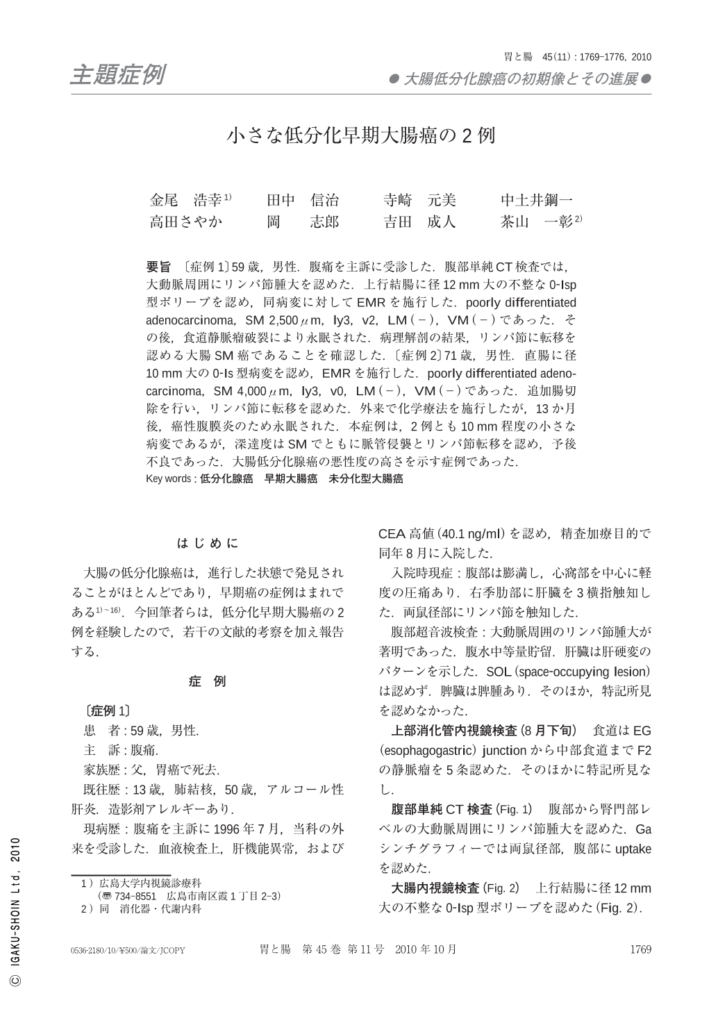
要旨 〔症例1〕59歳,男性.腹痛を主訴に受診した.腹部単純CT検査では,大動脈周囲にリンパ節腫大を認めた.上行結腸に径12mm大の不整な0-Isp型ポリープを認め,同病変に対してEMRを施行した.poorly differentiated adenocarcinoma,SM 2,500μm,ly3,v2,LM(-),VM(-)であった.その後,食道静脈瘤破裂により永眠された.病理解剖の結果,リンパ節に転移を認める大腸SM癌であることを確認した.〔症例2〕71歳,男性.直腸に径10mm大の0-Is型病変を認め,EMRを施行した.poorly differentiated adenocarcinoma,SM 4,000μm,ly3,v0,LM(-),VM(-)であった.追加腸切除を行い,リンパ節に転移を認めた.外来で化学療法を施行したが,13か月後,癌性腹膜炎のため永眠された.本症例は,2例とも10mm程度の小さな病変であるが,深達度はSMでともに脈管侵襲とリンパ節転移を認め,予後不良であった.大腸低分化腺癌の悪性度の高さを示す症例であった.
〔Case 1〕A 59-year-old-man. Abdominal CT revealed lymphnode swelling around the para-aorta. Colonoscopy revealed a type 0-Isp polyp in the ascending colon. It was 12mm in diameter. We performed EMR. Histological diagnosis was as follows : poorly differentiated adenocarcinoma ; with a depth of SM(2,500μm). The ly3, v2, lateral margin and vertical margin were negative. This patient died due to esophageal variceal rupture. As a result of the pathological autopsy, it was confirmed that this lesion was a colon submucosal cancer with lymph-node metastasis. 〔Case 2〕A 71-year-old-man. Colonoscopy revealed a type 0-Is polyp in the rectum, 10mm in diameter. We performed EMR. Histological diagnosis was as follows : poorly differentiated adenocarcinoma depth SM(4,000μm). The ly3, v0, lateral margin and the vertical margin were negative." Surgical resection was performed, and there was lymph node metastasis. This patient died of the peritonitis carcinomatosa 13 months later, despite the performance of chemotherapy. These cases showed the high malignant potential of poorly differentiated adenocarcinoma of the colon.
Copyright © 2010, Igaku-Shoin Ltd. All rights reserved.