Japanese
English
特集 腹部の最新画像情報2018
症例
強い濃染を呈した肝エキノコックス症の1例
Alveolar echinococcosis with strong enhancement in the liver
児玉 芳尚
1
,
櫻井 康雄
1
,
吉野 裕紀
1
,
古賀 英彬
2
,
真口 宏介
2
,
高田 実
3
,
大森 優子
4
Yoshihisa Kodama
1
1手稲渓仁会病院 放射線診断科
2同 消化器内科
3同 外科
4同 病理診断科
1Department of Radiology Teine Keijinkai Hospital
キーワード:
alveolar echinococcosis
,
肝癌
,
intrahepatic cholangiocarcinoma
,
強染
Keyword:
alveolar echinococcosis
,
肝癌
,
intrahepatic cholangiocarcinoma
,
強染
pp.531-536
発行日 2018年5月10日
Published Date 2018/5/10
DOI https://doi.org/10.18888/rp.0000000427
- 有料閲覧
- Abstract 文献概要
- 1ページ目 Look Inside
- 参考文献 Reference
- サイト内被引用 Cited by
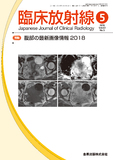
肝エキノコックス症は我が国では北海道に多い寄生虫疾患である。特徴的な画像を呈するため,比較的診断は容易であり,術前診断されることが多い疾患である。今回我々は,強く濃染し,肝内胆管癌との鑑別が困難であった肝エキノコックス症の1例を報告する。
A 61 year-old woman had backpain. US revealed low echoic mass with unclear margin in the liver. CT showed the tumor with strong heterogenous enhancement with bile duct dilation. MR showed low intensity on T1WI, and heterogenous high intensity with small cyst and bile duct dilation on T2WI. Intrahepatic cholangiocarcinoma was suspected because of enhancement pattern and bile duct dilation. Left hepatectomy was done. Final diagnosis was alveolar echinococcosis with massive inflammation in the liver.

Copyright © 2018, KANEHARA SHUPPAN Co.LTD. All rights reserved.


