Japanese
English
- 有料閲覧
- Abstract 文献概要
- 1ページ目 Look Inside
- 参考文献 Reference
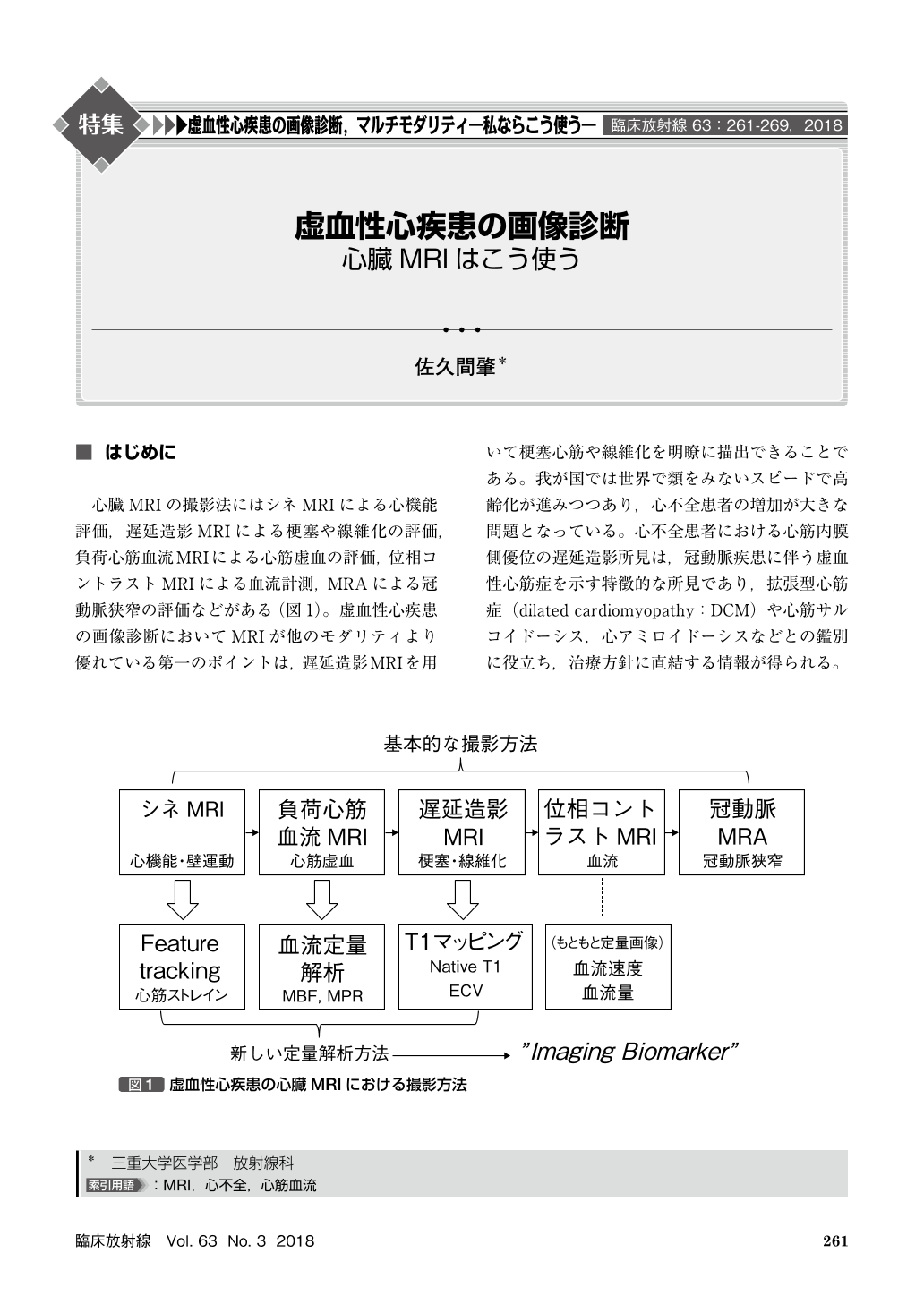
心臓MRIの撮影法にはシネMRIによる心機能評価,遅延造影MRIによる梗塞や線維化の評価,負荷心筋血流MRIによる心筋虚血の評価,位相コントラストMRIによる血流計測,MRAによる冠動脈狭窄の評価などがある(図1)。虚血性心疾患の画像診断においてMRIが他のモダリティより優れている第一のポイントは,遅延造影MRIを用いて梗塞心筋や線維化を明瞭に描出できることである。我が国では世界で類をみないスピードで高齢化が進みつつあり,心不全患者の増加が大きな問題となっている。心不全患者における心筋内膜側優位の遅延造影所見は,冠動脈疾患に伴う虚血性心筋症を示す特徴的な所見であり,拡張型心筋症(dilated cardiomyopathy:DCM)や心筋サルコイドーシス,心アミロイドーシスなどとの鑑別に役立ち,治療方針に直結する情報が得られる。
Cardiac MRI(CMR)permits accurate assessment of myocardial ischemia as well as detection of myocardial infarction and fibrosis. The prevalence of heart failure is increasing in Japan. CMR with late gadolinium enhanced MRI can accurately differentiate ischemic cardiomyopathy and dilated cardiomyopathy, and allows for exclusion of cardiac sarcoidosis and amyloidosis. Recent studies demonstrated high diagnostic accuracy of stress CMR study including perfusion MRI as compared with stress myocardial perfusion SPECT. Stress perfusion MRI is also shown to be useful in risk stratification of the patients with suspected coronary artery disease.

Copyright © 2018, KANEHARA SHUPPAN Co.LTD. All rights reserved.


