Japanese
English
- 有料閲覧
- Abstract 文献概要
- 1ページ目 Look Inside
I.緒言
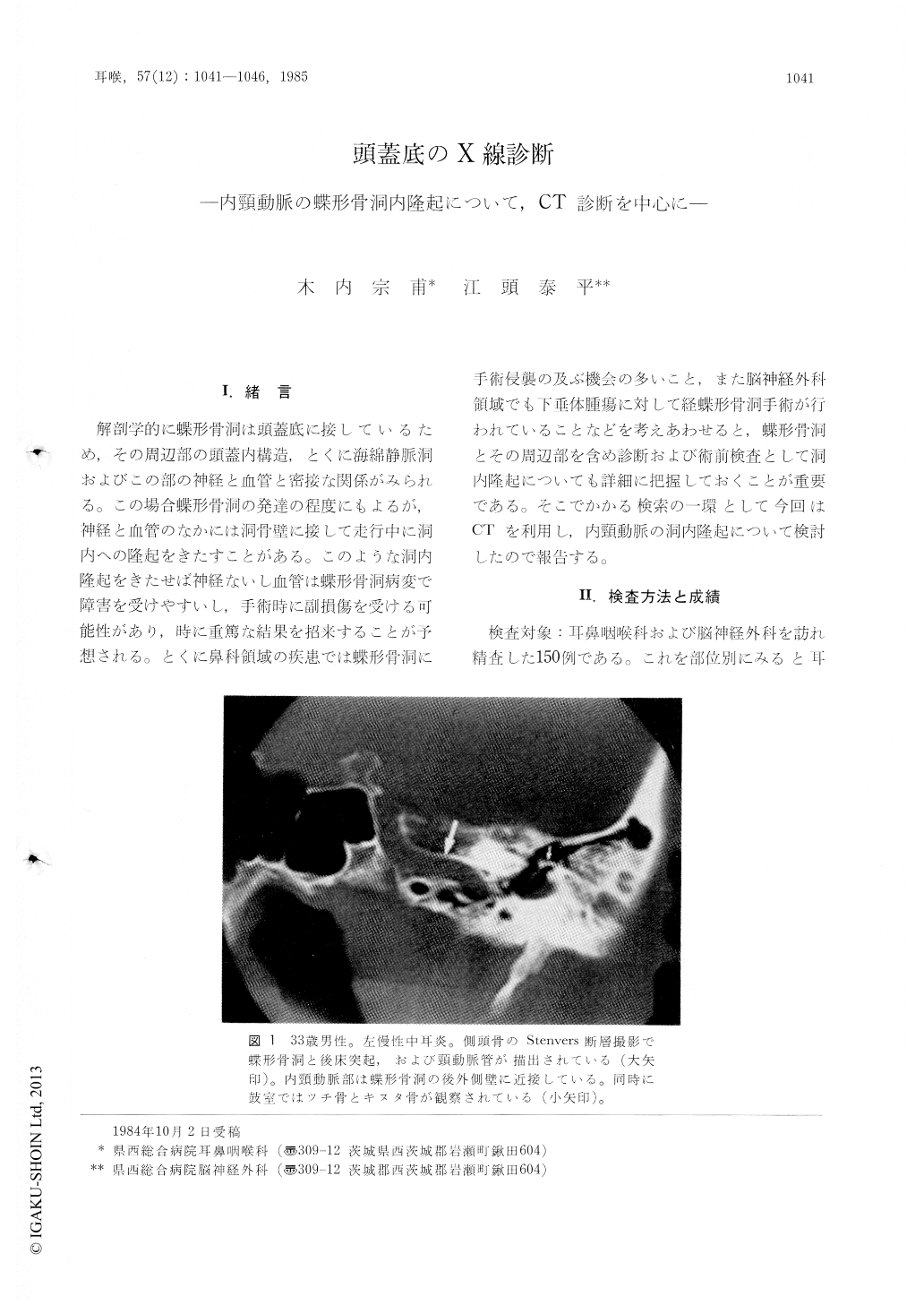
解剖学的に蝶形骨洞は頭蓋底に接しているため,その周辺部の頭蓋内構造,とくに海綿静脈洞およびこの部の神経と血管と密接な関係がみられる。この場合蝶形骨洞の発達の程度にもよるが,神経と血管のなかには洞骨壁に接して走行中に洞内への隆起をきたすことがある。このような洞内隆起をきたせば神経ないし血管は蝶形骨洞病変で障害を受けやすいし,手術時に副損傷を受ける可能性があり,時に重篤な結果を招来することが予想される。とくに鼻科領域の疾患では蝶形骨洞に手術侵襲の及ぶ機会の多いこと,また脳神経外科領域でも下垂体腫瘍に対して経蝶形骨洞手術が行われていることなどを考えあわせると,蝶形骨洞とその周辺部を含め診断および術前検査として洞内隆起についても詳細に把握しておくことが重要である。そこでかかる検索の一環として今回はCTを利用し,内頸動脈の洞内隆起について検討したので報告する。
To determine the frequency of the carotid prominence in the sphenoid sinuses, computed tomography was carried out in 150 patients with lesions mainly arised from the intracranial cavity, ear and paranasal sinuses. We classified the sphenoid sinuses into three types depending on the extent to which the sphenoid bone was pneumatized. In the first type the sphenoid sinuses did not extend into the body of the sphenoid bone. In the second type they extended more or less dorsally into the body of the sphenoid bone, and in the third type the entire floor of the sella was occupied by the sphenoid sinus. The second type was found in 22% and the third type in 77%.
In this series, the carotid prominence was observed in 62% of 109 patients, who showed the third type of the sphenoid sinuses, and the condition was prominent in 33%.

Copyright © 1985, Igaku-Shoin Ltd. All rights reserved.


