Japanese
English
- 有料閲覧
- Abstract 文献概要
- 1ページ目 Look Inside
I.はじめに
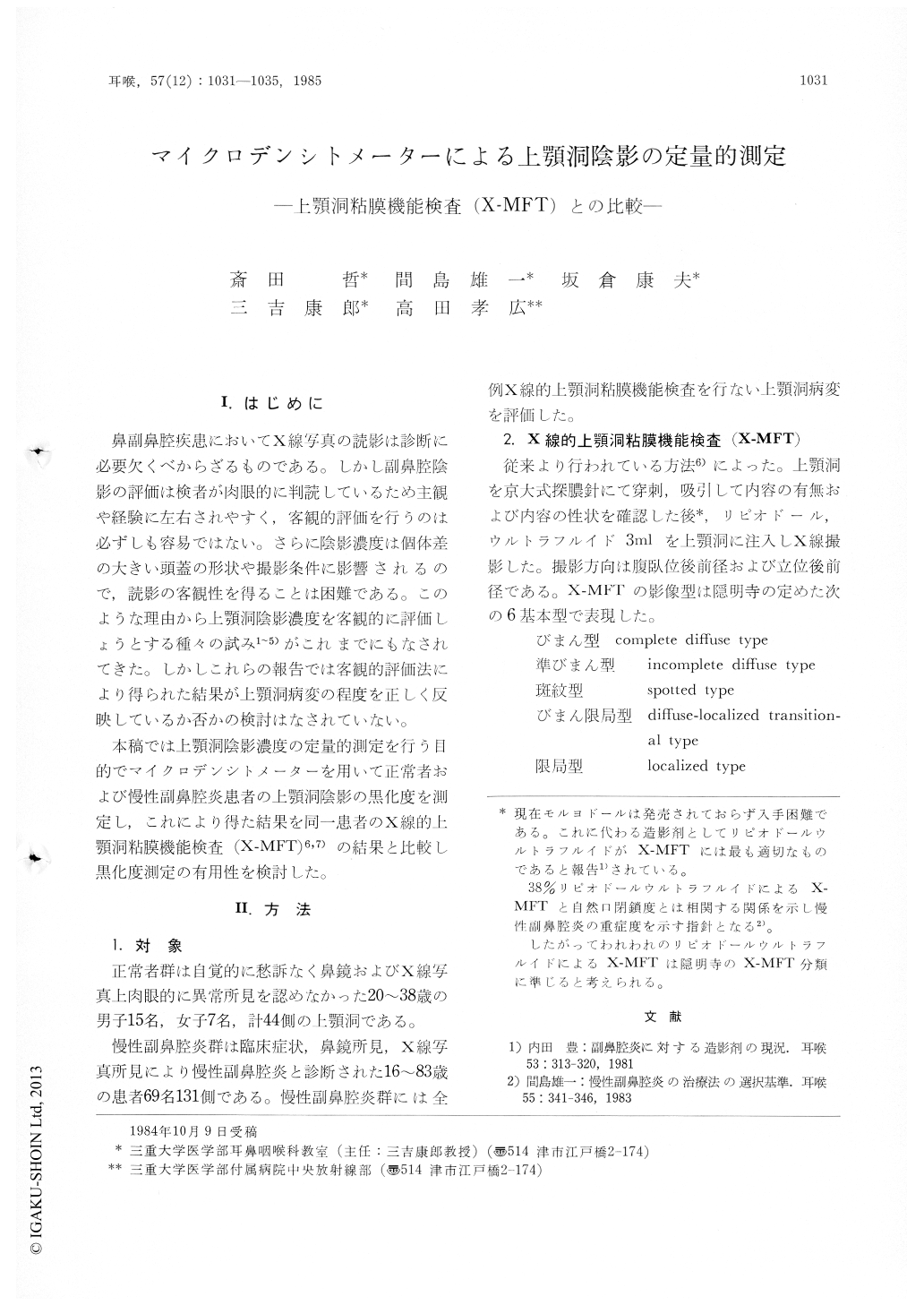
鼻副鼻腔疾患においてX線写真の読影は診断に必要欠くべからざるものである。しかし副鼻腔陰影の評価は検者が肉眼的に判読しているため主観や経験に左右されやすく,客観的評価を行うのは必ずしも容易ではない。さらに陰影濃度は個体差の大きい頭蓋の形状や撮影条件に影響されるので,読影の客観性を得ることは困難である。このような理由から上顎洞陰影濃度を客観的に評価しょうとする種々の試み1〜5)がこれまでにもなされてきた。しかしこれらの報告では客観的評価法により得られた結果が上顎洞病変の程度を正しく反映しているか否かの検討はなされていない。
本稿では上顎洞陰影濃度の定量的測定を行う目的でマイクロデンシトメーターを用いて正常者および慢性副鼻腔炎患者の上顎洞陰影の黒化度を測定し,これにより得た結果を同一患者のX線的上顎洞粘膜機能検査(X-MFT)6,7)の結果と比較し黒化度測定の有用性を検討した。
The opacity of the maxillary sinus on x-ray film (Waters view) was evaluated by microdensitometry in normal group and patients with chronic sinusitis. The ratio of the maxillary sinus density to the orbital density (M / 0 ratio) was calculated, and the results were compared among each group of subjects and between the groups.
The pathologic conditions of the maxillary sinus were classified into mild, moderate and severe lesions by x-ray mucous membrane function test (X-MFT). M / 0 ratio was significantly decreased as the severity of the maxillary lesion increased. Two sets of data were compared between pre- and post-treatment within a subject. Improved group on X-MFT was significantly improved on densitometric analysis, but unimproved group on X-MFT was unchanged.
These results indicated that evaluation of x-ray opacity of the maxillary sinus by microdensitometry was useful for quantitative evaluation of the pathologic condition of the maxillary sinus in chronic sinusitis.

Copyright © 1985, Igaku-Shoin Ltd. All rights reserved.


